JAMA & ARCHIVES
Arch Fam Med
SEARCH
GO TO ADVANCED SEARCH
HOME  PAST ISSUES  TOPIC COLLECTIONS  CME  PHYSICIAN JOBS  CONTACT US  HELP
Institution: CLOCKSS  | My Account | E-mail Alerts | Access Rights | Sign In
  Vol. 7 No. 2, March 1998 TABLE OF CONTENTS
  Archives
 • Online Features
 Article Links
 •Return to Article

Downloading the PowerPoint slide may take up to 30 seconds. If the slide opens in your browser, select File -> Save As to save it.
Copyright restrictions may apply. Please see our Conditions of Use.


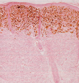
Figure 3. The upper dermis is occupied by heavily pigmented nevus cells that stained positive for HMB-45, while the lower dermis contains nonpigmented nevus cells that stained negative for HMB-45 (hematoxylin-eosin, x40).


HOME | CURRENT ISSUE | PAST ISSUES | TOPIC COLLECTIONS | CME | PHYSICIAN JOBS | HELP
CONDITIONS OF USE | PRIVACY POLICY | CONTACT US | SITE MAP
 
© 2011 American Medical Association. All Rights Reserved.

DCSIMG