|
|
Pathological Case of the Month
Gregg T. Lueder, MD, Contributor;
Bertram Matsumoto, MD, Contributor;
Morton E. Smith, MD, Contributor;
Enid Gilbert-Barness, MD, Section Editor
Arch Fam Med. 1998;7:207-208.
A 4-YEAR-OLD girl was referred for evaluation of a lesion of the left inferior conjunctiva that had been noticed 1 month earlier. Over the 2 days prior to her examination, small amounts of spontaneous hemorrhage had occurred from the lesion. Her medical history was unremarkable.
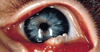
On examination, she had normal vision in both eyes. A 1 x 2-mm white lesion was present in the left inferior conjunctival fornix, with hairs appearing to arise from within the lesion (Figure 1). Results of the remainder of her ocular examination were normal.
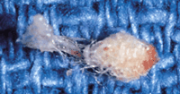
The patient was examined under anesthesia. The lesion was pedunculated, and the hairs on its surface were easily removed with a cotton-tip swab. The surrounding conjunctiva was erythematous, and follicles were present. With gentle manipulation, the lesion was peeled from the underlying conjunctiva. It appeared to contain synthetic fibers (Figure 2).
From the Departments of Ophthalmology and Visual Sciences (Drs Lueder, Matsumoto, and Smith), Pediatrics (Dr Lueder), and Pathology (Dr Smith), Washington University School of Medicine, St Louis Children's Hospital, St Louis, Mo.
|
Diagnosis and Discussion: Synthetic Fiber Granuloma (‘Teddy Bear Granuloma')
Figure 1. White lesion with adherent hairs in left inferior conjunctival fornix.
Figure 2. Lesion composed of tangled fibers that appear to be synthetic.
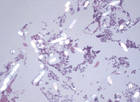
Figure 3. The lesion demonstrates areas of birefringence under examination with polarized light (hematoxylin-eosin, original magnification x40).
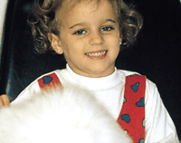
Figure 4. Patient with stuffed animal with long, synthetic hairs that can be easily removed.
Synthetic fiber granuloma ("teddy bear granuloma") of the conjunctiva was first described by Weinberg et al1 in 1984. The lesion results from an inflammatory reaction to synthetic fibers that become implanted in the conjunctival fornices (the space between the inner eyelid and the globe). In young children, these commonly result from close exposure to stuffed animals or blankets made of synthetic material. Affected patients present with ocular irritation secondary to the inflammation. The lesions are unilateral, and they are almost always found inferiorly. Eyelash hairs commonly adhere to the lesions.
Weinberg et al1 described several histologic features of synthetic fiber granulomas, which are used in their diagnosis. Gross examination reveals the presence of compressed, tangled fibers (Figure 2). Under the dissecting microscope, the ends of the fibers may demonstrate a light-pipe phenomenon. Hematoxylin-eosin staining of the fibers reveals a granulomatous inflammatory infiltrate, usually with foreign-body giant cells. The synthetic fibers do not have central cores, which are commonly seen in cilia and natural fibers. Scattered black granules may be seen within the synthetic material, resulting from titanium, barium, and zinc used in processing. Scanning electron microscopic examination of the fibers reveals an underlying smooth surface with longitudinal grooves. When the lesion is clinically suspected, the simplest method to confirm the diagnosis is the demonstration of marked birefringence when examined with polarized light (Figure 3).
The clinical differential diagnosis for this lesion includes an inflammatory reaction to other foreign bodies, such as hairs from the adjacent eyelashes or caterpillar hairs (conjunctivitis nodosa). The presence of a white lesion with hairs on the surface arising from the conjunctiva also suggests the diagnosis of a dermolipoma.
Removal of the lesion is required for successful treatment. Surgical excision is usually employed,1-3 although successful removal at the slitlamp with topical anesthesia has been reported in a 5-year-old child.4 If the lesion is recognized early, it is less likely to be encased in granulomatous tissue and adherent to the underlying conjunctiva. This would allow for removal in the office if the patient is able to cooperate during gentle manipulation at a slitlamp.5
After the nature of our patient's lesion was discussed with her mother, she reported that the child frequently slept with a large stuffed animal that shed fibers onto the surrounding bedding material (Figure 4). Contact with the teddy bear was eliminated, and the patient was treated with topical neomycin sulfate, polymyxin B sulfate, and dexamethasone ointment for 2 weeks. Three weeks after removal of the foreign body, the left inferior conjunctiva appeared to be normal.
AUTHOR INFORMATION
Selected from Arch Pediatr Adolesc Med. 1996;150:327-328. Pathological Case of the Month.
REFERENCES
|
1. Weinberg JC, Eagle RC, Font RL, Streeten BW, Hidayat A, Morris DA. Conjunctival synthetic fiber granuloma: a lesion that resembles conjunctivitis nodosa. Ophthalmology. 1984;91:867-872.
WEB OF SCIENCE
| PUBMED
2. Ferry AP. Synthetic fiber granuloma: ‘teddy bear' granuloma of the conjunctiva. Arch Ophthalmol. 1994;112:1339-1341.
FREE FULL TEXT
3. Shields JA, Augsburger JJ, Stechschulte J, Repka M. Synthetic fiber granuloma of the conjunctiva. Am J Ophthalmol. 1985;99:598-600.
4. Resnick SC, Schainker BA, Ortiz JM. Conjunctival synthetic and nonsynthetic fiber granulomas. Cornea. 1991;10:59-62.
FULL TEXT
|
WEB OF SCIENCE
| PUBMED
5. Lueder GT, Matsumoto B. Synthetic fiber granuloma. Arch Ophthalmol. 1995;113:848-849.
FREE FULL TEXT
|