|
|
Ischemic Scalp Necrosis Preceding Loss of Visual Acuity in Giant Cell Arteritis
Jay C. Rudd, MD;
Mitchell S. Fineman, MD;
Robert C. Sergott
Arch Fam Med. 1999;8:474-475.
GIANT CELL arteritis (GCA) is a systemic, necrotizing vasculitis affecting medium- to large-sized arteries. The most frequently involved arteries are the superficial temporal, vertebral, ophthalmic, and posterior ciliary.1-2 The incidence of GCA has been reported to be as high as 25 per 100,000 patients.3 Familiarity with the many systemic manifestations of GCA is essential for ophthalmologists.
As the most common cutaneous manifestation,4 scalp necrosis is well documented in the English-language medical literature5; however, GCA with associated scalp necrosis has rarely been reported in the ophthalmic literature. To alert ophthalmologists to this uncommon clinical finding, we describe a patient with acute loss of visual acuity due to biopsy-proven GCA preceded by ischemic scalp necrosis.
REPORT OF A CASE
An 85-year-old man was first seen with acute, painless, monocular loss of visual acuity in his right eye of 2 days' duration. He had developed an undiagnosed rash on his forehead that his dermatologist treated with bacitracin ointment 2 weeks prior to the loss of visual acuity. His symptoms included the following: mild headache, 9.0-kg weight loss, and jaw claudication.
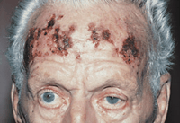
An erythematous, excoriated scalp lesion was present on the forehead in the distribution of both right and left superficial temporal arteries (Figure 1). Visual acuity was light perception OD and 20/40 OS. The temporal arteries were firm and cordlike bilaterally, with no palpable pulses. A marked right, relative afferent, pupillary defect was present. Visual fields were full by confrontation testing in the left eye. The right optic nerve head was diffusely edematous with associated flame hemorrhages. The left fundus showed no abnormalities.
|
|
|
|
Figure 1. Scalp lesions with eschars formed over an erythematous base, with lesions crossing the midline; no vesicles noted.
|
|
|
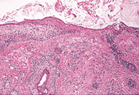
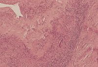
Intravenous methylprednisolone at 250 mg was instituted every 6 hours for presumed GCA. A Westergren erythrocyte sedimentation rate was 100 mm/h (reference range, 0-20 mm/h). A punch biopsy specimen of the skin showed focal epidermal necrosis and a sparse mononuclear infiltrate with necrosis of the substantia propria (Figure 2). Viral cultures were negative for growth. A temporal artery biopsy specimen confirmed the diagnosis of GCA (Figure 3).
|
|
|
|
Figure 2. Biopsy specimen of scalp lesions shows necrosis and ulceration of epidermic and foci of chronic inflammatory cells in the necrotic dermis (hematoxylin-eosin, original magnification x25).
|
|
|
|
|
|
|
Figure 3. Positive temporal artery biopsy specimen shows thickened intime, a mononuclear infiltrate, and giant cells near the internal elastic lamina (hematoxylin-eosin, original magnification x15).
|
|
|
The scalp lesions began to improve after 3 days of intravenous corticosteroids. Visual acuity remained light perception OD and 20/40 OS. The patient was discharged from the hospital and a regimen of oral prednisone and bacitracin ointment was started.
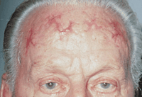
Visual acuity was unchanged 1 week later, and the scalp lesions continued to show improvement on examination 3 months after his first visit (Figure 4).
|
|
|
|
Figure 4. Three months after treatment, the scalp lesions have cleared, resulting in an erythematous scar.
|
|
|
COMMENT
Cutaneous involvement in GCA is well documented. Currey5 reviewed the English-language literature and found 24 cases of scalp necrosis associated with GCA; 16 patients had loss of visual acuity and 13 patients had bilateral scalp necrosis. The necrosis was most often distributed in the temporoparietal region. Findings from skin biopsies performed in the few previously reported cases did not demonstrate conclusive findings of GCA-associated skin necrosis. In our patient, a skin biopsy specimen revealed focal areas of necrosis and mononuclear cell infiltration.
In a review of 14 reported cases, Soderstrom and Seehafer6 noted that 67% of patients with GCA and scalp necrosis suffered irreversible loss of visual acuity. These patients also had a significant mortality rate (40%), most due to cardiopulmonary disease. The higher rate of loss of visual acuity and mortality in patients with GCA and scalp necrosis may reflect an aggressive vasculitis that is capable of completely obstructing the blood supply to the richly vascularized scalp. It may also represent a lack of awareness of the possible clinical significance of the dermatologic findings. In patients older than 55 years, scalp necrosis should prompt immediate evaluation for GCA.
AUTHOR INFORMATION
Selected from Arch Ophthalmol. 1998;116:1690-1691. Photo Essay.
REFERENCES
|
1. Keltner JL. Giant-cell arteritis: signs and symptoms. Ophthalmology. 1982;89:1101-1110.
ISI
| PUBMED
2. Goodman BW Jr. Temporal arteritis. Am J Med. 1979;67:839-852.
FULL TEXT
|
ISI
| PUBMED
3. Ghanchi FD, Dutton GN. Current concepts in giant cell (temporal) arteritis. Surv Ophthalmol. 1997;42:99-123.
FULL TEXT
|
ISI
| PUBMED
4. Baum EW, Sams WM Jr, Payne RR. Giant cell arteritis: a systemic disease with rare cutaneous manifestations. J Am Acad Dermatol. 1982;6:1081-1088.
ISI
| PUBMED
5. Currey J. Scalp necrosis in giant cell arteritis and review of the literature. Br J Rheumatol. 1997;36:814-816.
FREE FULL TEXT
6. Soderstrom CW, Seehafer JR. Bilateral scalp necrosis in temporal arteritis: a rare complication of Horton's disease. Am J Med. 1976;61:541-546.
FULL TEXT
|
ISI
| PUBMED
|