|
|
A Nonhealing Ulcer on the Face
Diana K. Sun, MD;
Daniel M. Siegel, MD
Arch Fam Med. 2000;9:787-789.
REPORT OF A CASE
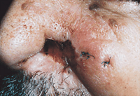
A 70-year-old Italian man presented with a 6-month history of a deep ulcerative lesion involving his left nasal ala after use of a nasal canula during a recent hospitalization. The lesion failed to heal and gradually expanded. At presentation, the patient was noted to be missing approximately half of the left nasal ala. In its stead, there was erosion surrounded by hemorrhagic crusting and an approximately 4-cm margin of mild erythema and edema over the left cheek. The erosion was indurated but nontender. At the left angle of the mouth, a 1.5 x 1.0-cm slightly waxy plaque with focal crusting was found. The nares appeared to have some mucosal inflammation. No lymphadenopathy was present (Figure 1).
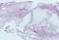
Four biopsy specimens were obtained: 2 punch biopsy specimens from the inflamed left cheek, and curettage biopsy specimens from the remaining alar border and the left angle of the mouth. The histopathologic findings are shown in Figure 2. Grocott, periodic acid–Schiff, and acid-fast bacillus stains were negative for organisms, as were fungal and acid-fast bacillus cultures. Gram stains of biopsy specimens from the left angle of the mouth and the left cheek were positive for cocci in clusters, consistent with Staphylococcus aureus.
From the State University of New York at Stony Brook.
|
Diagnosis and Discussion: Dermatitis artefacta
HISTOPATHOLOGIC FINDINGS AND CLINICAL COURSE
Histopathologic evaluation revealed granulation tissue and fibrinous exudate. There were acute and chronic inflammation, necrosis, foci of hemorrhage, and polarizable fibers.
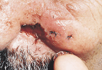
Given the histopathologic findings, the possibility of a trauma-induced lesion was raised. On questioning, the patient's family members recalled the patient applying a suction tube to his nose. The patient admitted to frequent scratching of his nose because of a "stuffed-up" sensation. He was found to have abundant cellular debris and dried blood under his left thumbnail (Figure 3).
DISCUSSION
Dermatitis artefacta is a condition in which the cutaneous lesions are self-inflicted and are the result or manifestation of some psychosocial conflict. Clinically, the lesions present in bizarre shapes, most commonly in a linear or geometric pattern.1-3 They are frequently related to the "handiness" of the individual, and are rarely in places that the patient cannot reach (unless an accomplice is involved). They tend to be clearly demarcated from the surrounding normal skin and appear on skin that was previously normal in appearance, often overnight. They do not evolve gradually, but rather emerge as fully developed lesions progressing toward healing. Therefore, they may be in different stages ranging from erythema, blisters, and denuded areas to crusts, pigmentary changes, and scars.2 The lesions of dermatitis artefacta may be produced by a variety of mechanical or chemical means, including bare hands, sharp or blunt objects, lit cigarettes, or substances such as phenol, silver nitrate, acids, or alkalies.2, 4 Some patients have even been reported to self-inject feces, milk, diphtheria prophylactic, and lighter fluid.5-7 The substances used may leave a smell on the skin or traces of dribbling if the caustic substances were applied or injected carelessly.2
Patients with dermatitis artefacta often are women who tend to be emotionally immature or have psychological or interpersonal difficulty. The range of intelligence of the patients is wide.4 A significant proportion of those afflicted are members of the medical profession, especially nurses, technicians, and paramedics.8 On presentation, patients with dermatitis artefacta may provide a "hollow history." The lesions seems to appear without any prior signs or symptoms. The patients often are indifferent toward the lesions. Frequently, they are unable or unwilling to discuss the appearance of the eruption in its early stages. Instead, the history is tangential.2-3,9 Some patients have also been noted to provide a "melodramatic prophecy" in which they can forecast the time and location of the new lesions.2 However, efforts by the physician to pursue the historic details may be met with hostility. The suggestion of self-infliction is often vehemently denied by the patient and family members.1, 4
Artifactual disease is frequently regarded as a result of emotional release. Often, some secondary gain is obtained by the patient, who may exhibit indifference toward what appears to be an incapacitating symptom.1
The lesions of dermatitis artefacta may manifest in a variety of ways, some of which can be quite extreme. Frequently, the patients undergo extensive medical testing to rule out organic disease. Physicians may be hesitant to diagnose dermatitis artefacta because they fear that they might be missing organic disease or perhaps because they are unwilling to believe that intelligent and respectable patients could be deceiving them.
When dermatitis artefacta is suspected, direct confrontation should be avoided. Evaluation of the patient's emotional situation or stresses should be made, and psychiatric counseling, if necessary, should be given. Sedatives and antidepressant agents may also be prescribed. In a study by Sneddon and Sneddon,10 33 patients with artifactual disease were followed up for 22 years. Most of the patients had long histories of psychological illness; a few had family difficulties. Seventy percent of the patients studied recovered from their cutaneous lesions after resolution of the underlying situation.
AUTHOR INFORMATION
Selected from Arch Dermatol. 2000;136:113. Off-Center Fold.
REFERENCES
|
1. Koblenzer CS. Psychosomatic concepts in dermatology. Arch Dermatol. 1983;119:501-511.
FREE FULL TEXT
2. Lyell A. Cutaneous artifactual disease. J Am Acad Dermatol. 1979;1:391-407.
PUBMED
3. Ostlere LS, Harris D, Denton C, et al. Boxing-glove hand: an unusual presentation of dermatitis artefacta. J Am Acad Dermatol. 1993;28:120-122.
PUBMED
4. Medansky RS, Handler RM. Dermatopsychosomatics: classification, physiology, and therapeutic approaches. J Am Acad Dermatol. 1981;5:125-136.
PUBMED
5. Steinman R, Mendelson J, Portnoy J. Self-inoculation with milk as a cause of recurrent cellulitis. CMAJ. 1975;112:605-606.
ABSTRACT
6. Boulton-Jones JM, Sissons JGP, Naish PF, et al. Self-induced glomerulonephritis. BMJ. 1974;3:387-390.
7. Shepard GH, Sawyers JL. Management of infection from self-inoculation with lighter fluid and other foreign agents. Ann Surg. 1969;170:292-295.
PUBMED
8. Hawkings R, Jones KS, Sim M, et al. Deliberate disability. BMJ. 1956;1:361-367.
PUBMED
9. Gandy DT. The concept and clinical aspects of factitial dermatitis. South Med J. 1953;46:551-555.
PUBMED
10. Sneddon I, Sneddon J. Self-inflicted injury: a follow-up study of 43 patients. BMJ. 1975;3:527-530.
|