


Effects of Mucosal Thickness on the Stress Distribution and Denture Stability of Mandibular Implant-Supported Overdentures with Unsplinted Attachments In Vitro
- Asuka Haruta1
- Yasuyuki Matsushita1
- Yoshihiro Tsukiyama1
- Yoshinori Sawae2
- Nobuo Sakai2
- Kiyoshi Koyano1
- 1Division of Oral Rehabilitation, Faculty of Dental Science, Kyushu University, 3-1-1 Maidashi, Higashi-ku, Fukuoka 812-8582, Japan
- 2Department of Intelligent Machinery and Systems, Faculty of Engineering, Kyushu University, 744 Motooka, Nishi-ku, Fukuoka 819-0395, Japan
- Asuka Haruta, aharuta{at}dent.kyushu-u.ac.jp
Abstract
The aim of this study was to compare the effects of mucosal thickness on the stress pattern around implants and movement of implant-supported overdentures with ball/female and three different types of magnetic attachments. After insertion of two root-form implants into a mandibular model, the surface of the model was covered with a 1.5- or 3-mm layer of impression material to simulate the oral mucosa, and removable overdentures were fabricated on each model. A 50-N vertical force was applied to the right first molar, and the resultant stress distribution and denture movement were measured. In the 1.5-mm mucosal model, the magnetic attachments showed significantly lower bending moments than did the ball attachment. The denture base displacement was the lowest on a magnetic attachment. In this study, use of magnetic attachments could be advantageous for mandibular implant-supported overdentures based on lower stress and better denture stability especially in the thin mucosal model.
1. Introduction
Implant-supported overdentures are useful for the treatment of compromised mandibular edentulous patients. Owing to their simplicity, comparatively low cost, and similar efficiency to fixed implant-supported prostheses, mandibular two-implant-supported overdentures have been considered by some to be the standard care for edentulous patients [1]. Several studies have indicated the clinical advantages of two-implant-supported overdentures in terms of retention, stability, and patient satisfaction [2–5].
Many different retention systems for overdentures have been proposed and used. Two-implant-supported overdentures on splinted implants with bar attachments or unsplinted implants with ball or magnetic attachments have shown high levels of clinical success [6]. Some in vitro studies have indicated that implant-supported overdentures on unsplinted implants show lower stress around the implants than do those on splinted implants [3, 7, 8].
Magnetic attachments have been applied as retention systems since the 1950s and are widely used in both natural teeth and dental implants [9, 10]. One of the greatest advantages of magnetic attachments is their reduced lateral forces, since lateral forces can badly influence the supporting teeth or implants. However, some clinical studies have shown that the retentive forces of magnetic attachments are significantly lower than those of bar or ball attachments [5, 11]. In addition, the designs of magnetic attachments have been associated with a number of problems including corrosion, wear, and demagnetization. Consequently, their designs have been improved against corrosion by using laser-welded coatings and their shapes have been altered to prevent elimination from the alveolar ridge, and the improved attachments have become additional available options for use with implant-supported overdentures. New magnetic attachments with a dome shape and soft material have been developed as stress distributors to allow displacement or rotation of the denture base during function, but their biomechanical characteristics have not been clarified because they have only been investigated in a few studies.
Implant-supported overdentures depend on support from both the implants and oral mucosa. The mucosal thickness of the alveolar ridge of the mandible was observed to be 2.0 mm on average, with a range of 0.6 to 4.8 mm [12]. A previous report [13] showed that denture movements caused by the different displacements of the implants and soft tissue caused stress accumulation around the implants. The mucosal thickness may affect the denture base displacement during function; therefore, the mucosal thickness probably influences the stress distribution. Some previous in vitro studies have indicated the effects of differences in the retentive systems for supported implants with spuriously soft tissue [8, 14], but these experiments were performed with only one mucosal thickness.
The purpose of this in vitro study was to compare the effects of mucosal thickness on the stress distribution around implants and on the movement of implant-supported overdentures with a ball attachment or three different types of magnetic retention systems.
2. Materials and Methods
2.1. Experimental Mandibular Model
An edentulous mandibular acrylic resin model (Palapress; Heraeus Kulzer, Germany) was fabricated. Two implants (4.1 mm in diameter, 12.0 mm in length; Straumann, Switzerland) were placed bilaterally in the canine region vertical to the residual ridge. They were set at 22 mm apart, similar to the distance between two natural canines. The implants were retained using resin cement (SuperBond CB; Sun Medical, Japan) (Figure 1). A 1.5-mm layer was removed from the denture-supporting surface of the resin model and replaced with polyvinyl siloxane impression material (Exafine injection type; GC, Japan) to simulate the resilient edentulous ridge mucosa. An experimental acrylic resin denture was conventionally fabricated on the model. In the same way, a 3-mm layer of impression material and a denture were fabricated on the same mandibular model. All experiments for the four attachments were carried out with one model of each mucosal thickness.
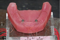
Edentulous mandibular model with implants placed in the canine region.
2.2. Measuring Devices
Four strain gauges (KFR-05-120-C-11; Kyowa Electronic Instruments, Japan) were attached to the mesiodistal and buccolingual sides of the neck part of each implant to measure the strain on the implants (Figure 2). The electric signals from the strain gauges were amplified, transmitted, and recovered by a personal computer (Aptiva 2168-S65; IBM, Japan) following A/D conversion (PCD-200A; Kyowa Electronic Instruments, Japan).
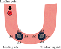
Measuring devices. Four strain gauges were attached to each implant.
A movement sensor (3SPACE; Polhemus, USA) was placed on the left first molar region to measure the displacement of the denture (Figure 3). The sensor used electromagnetic fields to determine the position and orientation of a remote object. The output of the movement sensor was input into a computer (OptiPlex GX1; Dell, Japan), and a mathematic algorithm calculated the position of the receiver relative to the transmitter and recorded the results. Meanwhile, denture movement at the loading side (right first molar region) was measured by vertical displacement of Autograph (AGS-10kng; Shimadzu, Japan) (Figure 3).
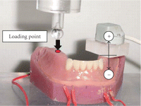
Experimental model with artificial mucosa and denture base. A movement sensor was placed to the left first molar region opposite the loading point.
2.3. Attachments
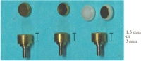
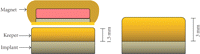
Prefabricated ball and magnetic attachments were used to attach the denture to the implants. The ball attachment consisted of an anchor head (048.439; Straumann, Switzerland) and a metal female component (048.456; Straumann, Switzerland) (Figure 4). Three different shapes of magnetic attachments were used: flat type (IP-DXFL; Aichi Steel Co., Japan), dome-shaped type (IP-MCD; Aichi Steel Co., Japan), and cushion type (IP-MCS; Aichi Steel Co., Japan) (Figure 5). The flat type was a typical magnetic attachment, while the dome-shaped type had a dome-shaped surface of the magnet and keeper, and the cushion type had a stress distributor with a magnet (Figure 6). The magnetic attachments consisted of a keeper as the magnet head and a magnet that was embedded in the denture. The keeper was prefabricated and had threads that were identical to the threads of an abutment screw. The heights of the keepers were 1.5 and 3 mm (Figure 7).
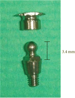
Ball attachment.
Magnetic attachments.
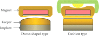
Dome-shaped type with a dome-shaped surface of the magnet and keeper and cushion type with a stress distributor.
The heights of the magnetic attachments.
2.4. Experiments
Autograph applied a load to the occlusal surface of the right first molar region. This study used a one-point concentrated load on the molar part that was considered to receive the load with the largest force during function. Loads from 0 to 50 N were applied gradually to simulate a moderate level of biting force on an implant-retained overdenture [8]. Each experiment was repeated five times under the same conditions. Each sequence of strain data was used to calculate the axial force and the bending moment transmitted to the implant [15]. The data were expressed as mean and standard deviations. Statistical comparisons were carried out using two-way analysis of variance (P < .05) and t-tests with the Bonferroni correction (SPSS ver. 17; SPSS Inc., Chicago, IlL, USA).
3. Results
3.1. Axial Force on the Implants
The axial force on the implants is shown in Figure 8. The plus-force showed tensile stress and the minus-force showed compressed stress. In the 1.5-mm mucosal model, the ball attachment showed a significantly higher axial force on the implants than all the magnetic attachments at both the loading and non-loading sides. On the other hand, in the 3-mm mucosal model, the ball attachment had a lower vertical force than all the magnetic attachments at the loading side and had a tensile stress at the non-loading side. For the 3-mm mucosal model, the dome-shaped type caused the highest axial force at the loading side. There were no significant differences among the three magnetic attachments in the 3-mm mucosal model at the non-loading side.
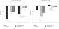
Axial force in the 1.5- and 3-mm mucosal models at the loading side and non-loading side. *p < .05.
3.2. Bending Moment on the Implants
The bending moment on the implants is shown in Figure 9. In the 1.5-mm mucosal model, all the magnetic attachments showed significantly lower bending moments on the implants than the ball attachment at both the loading and non-loading sides. In the 3-mm mucosal model, the bending moments of the magnetic attachments were higher than that of the ball attachment at the loading side. At the non-loading side, all the magnetic attachments showed lower bending moments than the ball attachment. The dome-shaped type caused the largest bending moment at the loading side, while it caused the lowest bending moment at the non-loading side. The flat type made a smaller bending moment than the dome-shaped type and cushion type at the loading side in each mucosal model.
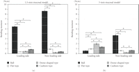
Bending moment in the 1.5- and 3-mm mucosal model at the loading side and non-loading side. *p < .05.
3.3. Denture Movement
The denture movement in the upward-downward direction is shown in Figure 10. The plus-movement showed upward displacement from the model, and the minus-movement showed downward displacement. In the 1.5-mm mucosal model, the denture base movements were larger on the ball attachment at both the loading and non-loading sides. At the loading side, the denture movement was the lowest on the flat type. On the other hand, at the non-loading side, the denture movement was very small on the magnetic attachments. In the 3-mm mucosal model, the denture base movement was larger on the cushion type. At the non-loading side on the 3-mm mucosal model, upward movement was shown on the magnetic attachments.
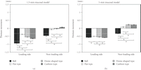
Denture movement in the 1.5- and 3-mm mucosal model at the loading side and non-loading side. *p < .05.
4. Discussion
The present in vitro study investigated the effects of differences in unsplinted attachments regarding the stress distribution and denture base displacement in models of different mucosal thicknesses.
The axial force and bending moment on the ball attachment were clearly larger in the 1.5-mm mucosal model than those in the 3-mm mucosal model. These findings are thought to be related to the height of the male part of the ball attachment, which acted as a lever. The height of this attachment’s male part was 3.4 mm. Consequently, the bending moment of the ball attachment was smaller in the 3-mm mucosal model than that in the 1.5-mm mucosal model because the height of the male part on the mucosa in the 3-mm mucosal model was smaller than that in the 1.5-mm mucosal model. On the other hand, the magnetic attachments had two heights at 1.5 and 3 mm, which suited the mucosal thickness in each model. However, the bending moment of the ball attachment was larger at the non-loading side, although the height of the ball attachment’s male part suited the 3-mm mucosal thickness. This was thought to be the effect of the retentive force on the ball attachment. The retentive force on the ball attachment was large, and the male and female parts of the ball attachment did not detach during testing. Therefore, the stress was transmitted to the implant at the non-loading side on the ball attachment. All of the magnetic attachments showed significantly lower bending moments than did the ball attachment at the non-loading sides.
The axial force of the ball attachment in the 3-mm mucosal model was a tensile force at the non-loading side. This meant that the denture base rotated around the connection of the male and female parts as a fulcrum. This was probably why the retentive force of the ball attachment was large.
Stress reportedly increased on the implants with a ball attachment as the thickness and resiliency of the mucosa increased [16]. This finding was different from that in the present study because of the height of the ball attachment and the resiliency of the mucosa. In this study, the effect of the difference in mucosal thickness and the resiliencies of 1.5- and 3-mm mucosa were identical because they were made of the same material. There was a possibility of different results for different resiliencies of the mucosa.
In the 1.5-mm mucosal model, the axial forces, bending moments, and denture base displacements were smaller on the magnetic attachments than those on the ball attachment. In the 1.5-mm mucosal model, the bending moments of all the magnetic attachments were sufficiently small, and the flat type caused the smallest denture base displacement. Therefore, the flat type is considered to be the most favorable attachment for this situation. In the 3-mm mucosal model, the axial forces on the magnetic attachments were larger those that on the ball attachment, and they transmitted the loading force to the implants axially. In the 3-mm mucosal model, the axial force and bending moment of the dome-shaped type at the loading side were the largest among all the attachments, because the contact between the magnet and the keeper was assumed to be maintained under this situation. The experiments were carried out under dry conditions, which probably caused larger friction.
Some previous studies have investigated the biomechanical characteristics of the magnetic attachments. A previous report [17] compared a stud type, an anchor type, and the three types of magnetic attachments for the lateral forces on the implants and the denture movement by in vitro experiments but did not find any significant differences among the magnetic attachments. On the other hand, the cushion type reportedly had little effect on the denture displacement and exhibited lower lateral stresses than did the flat type [14]. However, these studies were only performed with a 2-mm mucosal model. In the present study, we examined two different mucosal thicknesses and observed similar axial forces and bending moments with the flat type and cushion type, although the denture base movement was larger on the cushion type than that on the flat type. Generally, a larger denture base movement is associated with a larger bending moment on the implants. Therefore, these findings suggested that the cushion type was helpful for mitigating the lateral stress on the implants. These results probably indicate that the dome-shaped type and cushion type are more useful than the flat type owing to reduced wearing of their surfaces because they move out of place less frequently for each function. Further studies involving repetitive loading tests are required to clarify the wearing of these magnetic attachments.
In this study, vertical load was applied in each model with reference to a previous study [8]. It was thought that oblique load is more clinically related and harmful for stress distribution on the implants. However, obtaining stable outcomes was difficult; thus, vertical load was applied. The results showed that the magnetic attachments were useful for stress distribution and denture stability. However, the magnetic attachments were not stable when the dentures were subjected to lateral loads. It was possible to obtain different results by applying different load directions.
A good understanding of implant biomechanics makes it possible to optimize the treatment plan for individual patients to reduce the risk of functional complications and failures. Clinically, it can be hypothesized that the attachment systems providing the most equitable transfer of the occlusal forces among abutments are preferable from the standpoint of bone preservation.
5. Conclusions
Within the limits of this study, the findings indicated that the magnetic attachments were more favorable than the ball attachment in terms of the stress distribution and the denture base stability on a thin mucosa. For a thick mucosa, the flat type caused the smallest bending moment and denture base movement among all the attachments, suggesting that the flat type was the most favorable for this condition.
- Received December 9, 2010.
- Revision received March 1, 2011.
- Accepted March 15, 2011.
- © 2011 Asuka Haruta et al.


