- Institution: Stanford Univ Med Ctr Lane Med Lib/Periodical Dept/Rm L109
- Sign In as Member / Individual
PROMISCUOUS LIGANDS AND ATTRACTIVE CAVITIES
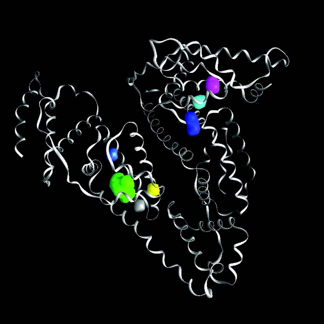
Figure 9.
Structure of human serum albumin [Protein Data Bank #1AO6 (40)], showing the carbon backbone as ribbons, and several internal cavities. These cavities are depicted as solids, representing the cavity surface, with color used for distinction. Only the green cavity is sufficiently large to bind a typical inhaled anesthetic without distortion of the native protein structure.

