- Institution: Stanford Univ Med Ctr Lane Med Lib/Periodical Dept/Rm L109
- Sign In as Member / Individual
Spinal Astrocytes in Pain Processing: Non-Neuronal Cells as Therapeutic Targets
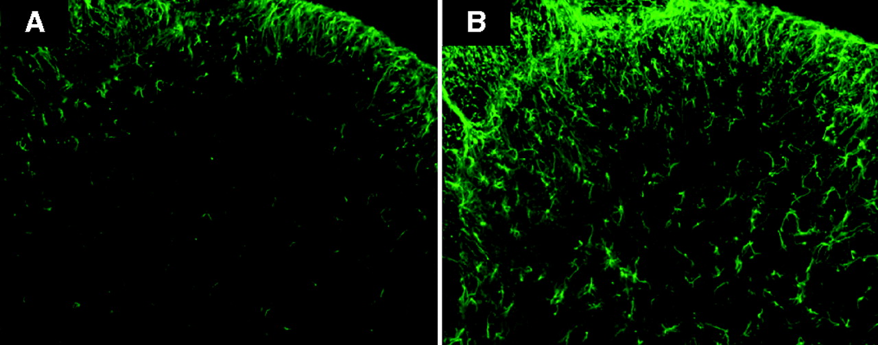
Figure 4
Immunohistochemistry images depicting GFAP immunoreactivity. A) Tissue from the naïve mouse dorsal horn is compared to B) tissue from an animal with collagen-antibody–induced arthritis. Note the increased GFAP signal intensity and astrocyte hypertorphy in the spinal dorsal horn of the animal with arthritis.

