- Institution: Stanford Univ Med Ctr Lane Med Lib/Periodical Dept/Rm L109
- Sign In as Member / Individual
Pulse-Chase in the Light Microscope
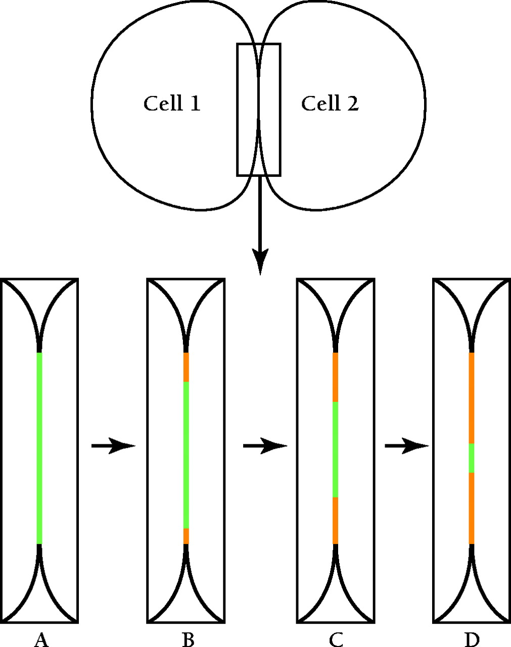
Figure 1.
A diagram summarizing the visualization of pulse–chase of connexin 43 (Cx43) in gap junctions. Two cells are diagrammed; the boxed area of cell–cell interaction is enlarged in panels A–D. (A) Cells transfected with tetracysteine-tagged Cx43 are labeled with FlAsH-EDT2, which stains the gap junction fluorescent green. Following a wash, to remove excess FlAsH-EDT2, cells are treated with ReAsH-EDT2 to label all newly synthesized Cx43 fluorescent red. (B–D) Over time, newly synthesized Cx43 (red) replaces pre-existing Cx43 (green) by accretion at the edges of the gap junction with concomitant removal of connexons from the center.

