- Institution: Stanford Univ Med Ctr Lane Med Lib/Periodical Dept/Rm L109
- Sign In as Member / Individual
Act Locally: New Ways of Regulating Voltage-Gated Ion Channels
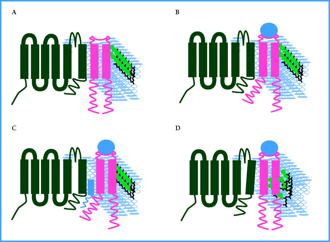
Putative models of sigma receptor modulation of ion channels. The plasma membrane is diagrammed to contain an idealized voltage-dependent ion channel (black) in close proximity to a sigma receptor (pink). The phopsholipid bilayer is depicted in blue and cholesterol molecules are green. When the unbound receptor and ion channel (as shown in A) binds to a ligand (blue circle), a conformational change occurs in the transmembrane or intracellular domains (or both) of the sigma receptor so that channel accessibility is either directly (as in B) or indirectly (as in C) regulated. Alternatively, ligand binding may result in local changes of cholesterol structure due to the activity of, or binding to, sterol isomerase-like sequences in the sigma receptors (D).

