- Institution: Stanford Univ Med Ctr Lane Med Lib/Periodical Dept/Rm L109
- Sign In as Member / Individual
BLOOD-BRAIN BARRIER DRUG TARGETING: THE FUTURE OF BRAIN DRUG DEVELOPMENT
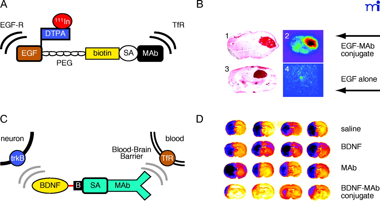
Delivery of protein therapeutics to the brain. A. Structure of an epidermal growth factor (EGF) chimeric peptide formed by conjugating the EGF to a molecular Trojan horse consisting of a monoclonal antibody (MAb) to the BBB transferrin receptor (TfR). See text for details. Thus, the EGF chimeric peptide is a bifunctional molecule that binds to the BBB TfR to allow for transport across the BBB, and to the EGF receptor (EGF-R) to allow for sequestration on the brain cancer cell membrane. Reprinted with permission (42). B. Panels 1 and 3 are autopsy sections of nude-rat brain bearing human U87 gliomas and stained with a MAb that binds the human EGF-R. The size of the tumor is visualized with the immunocytochemistry. Panels 2 and 4 are brain scans of the same rats as shown in Panels 1 and 3, but prior to sacrifice. The live nude rats bearing intracranial U87 human gliomas were administered intravenously either [111In]-EGF alone (Panel 4) or the [111In]-EGF-MAb chimeric peptide (Panel 2), indicating that EGF alone does not cross the BBB (Panel 4). However, the tumor is visualized with EGF chimeric peptide owing to transport of the EGF chimeric peptide across the BBB in the tumor (Panel 2). Reprinted with permission (42). C. Structure of a chimeric peptide of brain derived neurotrophic factor (BDNF) that is conjugated to a TfR MAb through an SA–biotin (B) linkage. The BDNF chimeric peptide is a bifunctional molecule that can bind both the BBB TfR to allow for transport from blood to brain, and the neuronal trkB receptor to allow for neuroprotection in brain. Reprinted with permission (47). D. Coronal sections of rat brain stained with 2,3,5-triphenyltetrazolium chloride (TTC). Coronal sections are shown for four different rats in four different treatment groups including saline, BDNF alone, TfRMAb alone, or the BDNF–TfR MAb chimeric peptide. The BDNF was administered at a dose of 50 μg/rat intravenously following permanent occlusion of the middle cerebral artery. The coronal slabs were scanned and the grayscale image was inverted and colorized so that the infarcted region appears dark purple and the healthy brain tissue appears yellow/red. There is no reduction in stroke volume with the BDNF alone because the neurotrophin does not cross the BBB, even in the infarcted region of brain. However, there was a 65% reduction in stroke volume following the delayed intravenous injection of the BDNF chimeric peptide because the neurotrophin was enabled to cross the BBB and enter into the ischemic brain region. Reprinted with permission (47).

