- Institution: Stanford Univ Med Ctr Lane Med Lib/Periodical Dept/Rm L109
- Sign In as Member / Individual
Galanin in Alzheimer Disease
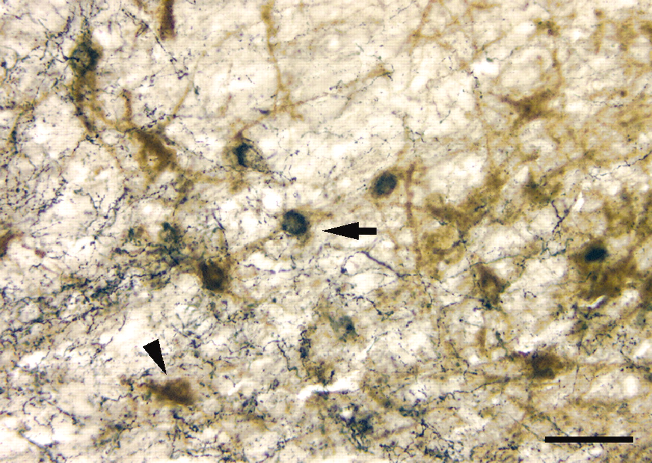
Figure 4.
GAL innervation of basal forebrain HDB following immunolesion with the selective cholinergic cell toxin 192IgG–saporin. Photomicrograph of ipsilateral lesioned HDB shows p75NTR-ir CBF neurons (brown reaction product; arrowhead) and p75NTR-ir CBF neurons containing heavy deposits of GAL immunoreactivity (dark blue reaction product; arrow). The beaded and punctate dark blue deposits resembling GAL-ir axons and terminals covering some of the p75NTR-ir dendrites are visible. Scale bar = 50 mm.

