- Institution: Stanford Univ Med Ctr Lane Med Lib/Periodical Dept/Rm L109
- Sign In as Member / Individual
Na+-K+–ATPase-Mediated Signal Transduction: From Protein Interaction to Cellular Function
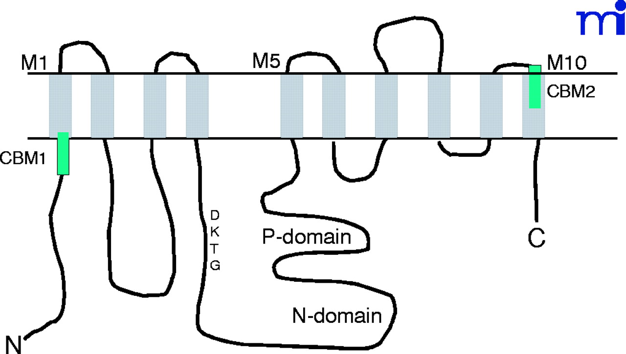
Figure 1.
Schematic presentation of the α1subunit of Na+-K+-ATPase. The ten transmembrane domains are marked as M1 to M10. The third intracellular loop, connecting the M4 and M5, contains both P (phosphorylation) domain and N (nucleotidebinding) domain. The green bars indicate caveolin-binding motifs (CBMs).

