- Institution: Stanford Univ Med Ctr Lane Med Lib/Periodical Dept/Rm L109
- Sign In as Member / Individual
Paradoxical Role of T-type Calcium Channels in Coronary Smooth Muscle
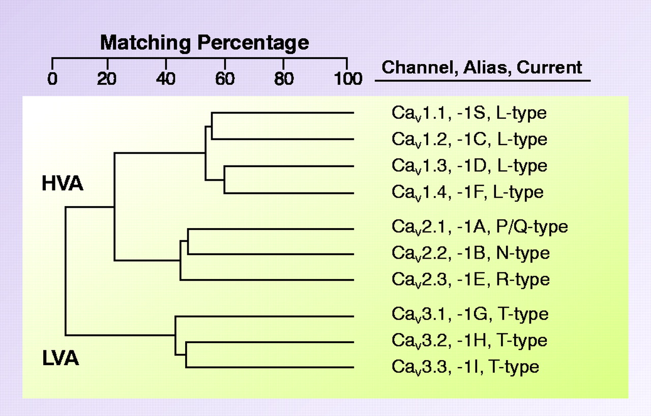
Family of voltage-gated calcium channels. Cloning of mammalian Ca2+ channels revealed the existence of ten genes. Alignment of their deduced amino acid sequences indicates that these channels can be grouped into three subfamilies, providing the basis for a rational nomenclature scheme. This family tree is based on the full-length human sequences, and differs from that obtained using membrane spanning regions (1). The Cav 1 channels carry L-type currents, and play critical roles in muscle excitation-contraction coupling. They are also the target of traditional “calcium channel blockers.” The Cav 2 channels are a second subfamily of high voltage–activated channels (HVA), and they play a dominant role in neurotransmitter release. Although subtype-selective small-molecule drugs have not been developed to for Cav 2 channels, these channels can be distinguished with peptide toxins. The Cav 3 channels carry T-type currents. These channels open after small depolarizations of the plasma membrane, and hence are also termed low voltage–activated (LVA) channels. This property allows Cav 3 channels to act as a pacemaker current, such as that originally observed in low threshold spiking and rebound burst firing of neurons (22).

