- Institution: Stanford Univ Med Ctr Lane Med Lib/Periodical Dept/Rm L109
- Sign In as Member / Individual
The Choline Transporter Resurfaces: New Roles for Synaptic Vesicles?
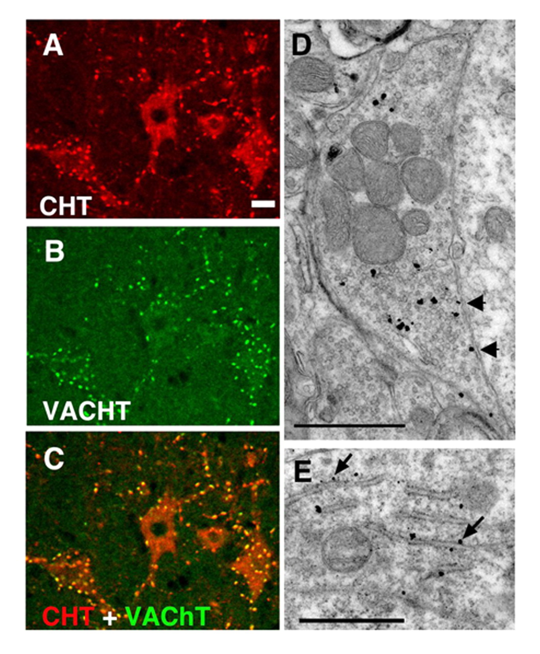
CHT colocalizes with VAChT at presynaptic terminals of cholinergic neurons. (A) In the ventral horn of the mouse spinal cord, CHT immunoreactivity is present in motor neuron cell bodies as well in the large presynaptic C-boutons that surround them (Scale bar = 20 μ m). (B) VAChT is present in the same cell bodies and terminals as CHT, confirming their cholinergic identity. (C) The distributions of CHT and VAChT overlap completely demonstrating that CHT is a constant feature of cholinergic neurons and that it is enriched at presynaptic sites. (D) Immuno-electron microscopy with CHT-specific antibodies illustrates that within an individual cholinergic presynaptic C-bouton, CHT immunoreactivity revealed by silver enhanced gold particles is predominantly associated with presynaptic vesicles and is only occasionally detected at the plasma membrane (arrows). Note that the epitope is on the cytosolic face of the plasma membrane, consistent with the predicted topology of CHT (scale bar = 1 μ m). (E) In the cell body of a cholinergic neuron, a biosynthetic pool of CHT immunoreactivity is labeled with the C-terminal epitope found on the cytoplasmic face of the rough endoplasmic reticulum (scale bar = 1 μ m). [Data adapted from (66).]

