- Institution: Stanford Univ Med Ctr Lane Med Lib/Periodical Dept/Rm L109
- Sign In as Member / Individual
Twenty Years of Calcium Imaging: Cell Physiology to Dye For
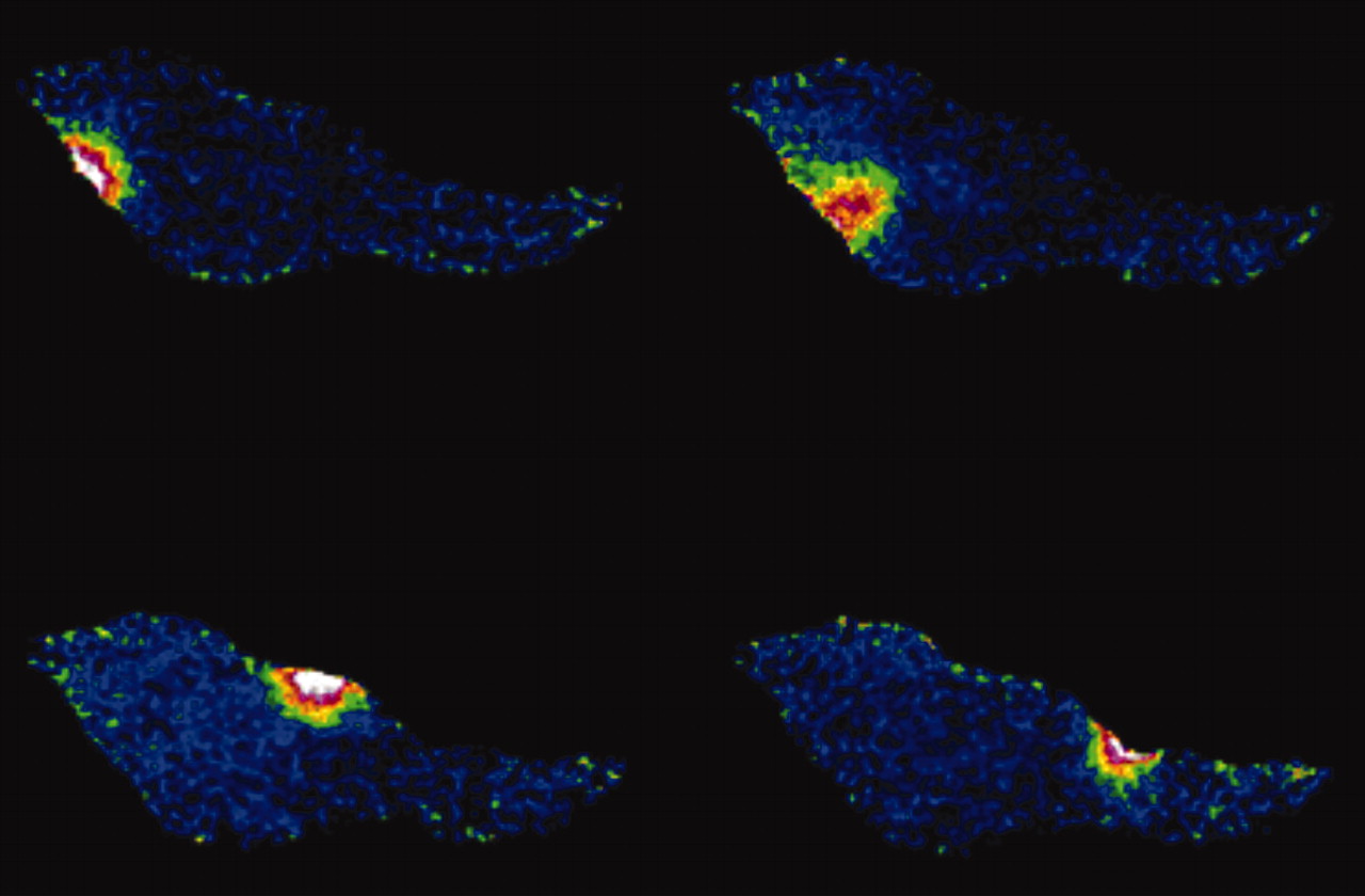
Figure 1.
Calcium sparks. These signals are spontaneous, transient, small bursts of sub-membrane Ca2+ originating from clusters of Ca2+-release channels on the sarcoplasmic reticulum (Ca2+ stores) as shown here for an isolated vascular smooth muscle cell at 4 time points (>500 ms apart).

