- Institution: Stanford Univ Med Ctr Lane Med Lib/Periodical Dept/Rm L109
- Sign In as Member / Individual
Twenty Years of Calcium Imaging: Cell Physiology to Dye For
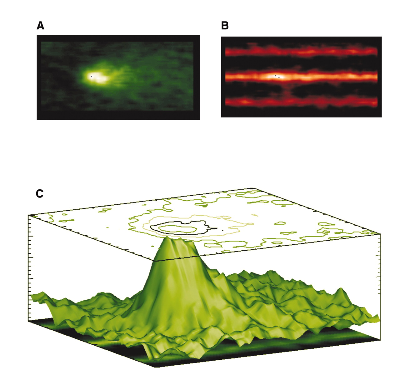
Figure 3.
Calcium sparks and transverse tubules (TTs). The Ca2+ indicator fluo-3 was loaded into heart cells to enable Ca2+ sparks to be visualized on a confocal microscope. A. Signal-averaged Ca2+ sparks. B. Sulforhodamine B was added to the extracellular solution to image the TTs, and Ca2+ sparks were imaged simultaneously. C. A surface plot shows the relationship between signal-averaged Ca2+ sparks (A) and TTs (B). The site of the origin of the majority of Ca2+ sparks is the TT from the junctional SR (jSR). [See (29).]

