- Institution: Stanford Univ Med Ctr Lane Med Lib/Periodical Dept/Rm L109
- Sign In as Member / Individual
Twenty Years of Calcium Imaging: Cell Physiology to Dye For
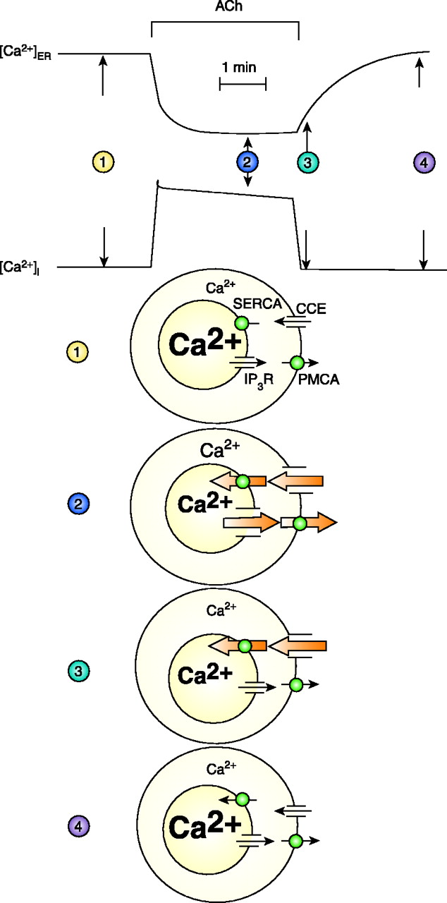
Overall Ca2+homeostasis in pancreatic acinar cell. The top two traces show [Ca2+] in the lumen of the ER and the cytosol, before, during, and after maximal ACh stimulation. Below are shown representations of the state of the four principal Ca2+ transporters in four situations: 1) Rest, before start of stimulation. Small fluxes of Ca2+ through Ca2+ release channels in the ER (for simplicity only the IP3 receptor is shown), ER Ca2+ pumps (SERCA), the plasma membrane Ca2+ pump (PMCA), and the store-operated Ca2+ entry pathway (also known as capacitative Ca2+ entry; CCE). Fluxes across both plasma and ER membranes are in equilibrium. 2) Steady-state stimulated situation. After the initial release of Ca2+ from the ER, both Ca2+ pumps (SERCA and PMCA) are now fully activated and the store-operated Ca2+ entry path is also open, due to the reduced [Ca2+]ER. 3) Immediately after cessation of ACh stimulation. The IP3 receptor channels have closed and the SERCA pump can now refill the ER via Ca2+ entering through the plasma membrane. The PMCA is no longer activated because [Ca2+]i has quickly returned to the basal level. 4) Normal Ca2+ gradients have been restored to the resting basal level. [Adapted from (90).]

