- Institution: Stanford Univ Med Ctr Lane Med Lib/Periodical Dept/Rm L109
- Sign In as Member / Individual
Twenty Years of Calcium Imaging: Cell Physiology to Dye For
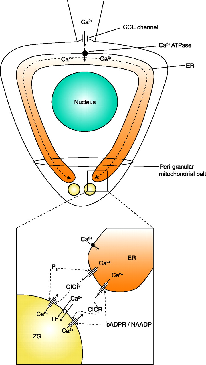
Ca2+signal generation in the apical granular pole. The main (upper) part shows a schematic diagram of a pancreatic acinar cell with the base at the top and the apical pole at the bottom. Ca2+ entry is depicted from a point source (cell-attached patch pipette) at the base. Ca2+ entering (through capacitative Ca2+ entry channels; CCE) is taken up by the ER Ca2+ pump and diffuses in the lumen of the ER towards the apex. At the apical pole, Ca2+ can be released through IP3 receptors and ryanodine channels, which can be stimulated, respectively, by IP3 and cADPR or NAADP (acting via different intermediary receptor proteins, but both ultimately activating the ryanodine receptor). Ca2+ released from the ER terminals can induce further Ca2+ release (CICR) from the secretory (zymogen granules; ZG), assisted by IP3 and cADPR/NAADP. The cytosolic Ca2+ signals are confined to the apical pole by the peri-granular mitochondrial belt. [Adapted and up-dated from (104).]

