- Institution: Stanford Univ Med Ctr Lane Med Lib/Periodical Dept/Rm L109
- Sign In as Member / Individual
MOSAICISM OF THE RETINAL PIGMENT EPITHELIUM: seeing the small picture
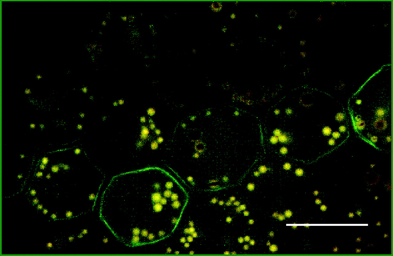
Figure 2.
Whole mount of the bovine RPE monolayer immunostained for the intermediate filament protein vimentin to illustrate a mosaic pattern of protein expression. Vimentin has a circumferential distribution in the peripheral cytoplasm (green) within a row-like subset of RPE cells. The tissue shown here is from the tapetal region of the cow eye which has relatively few melanosomes (brown granules), lipofuscin (yellow granules), and combined melanolipofuscin granules. Specimen was prepared as described in Figure 1. Scale bar: 20 μm.

