- Institution: Stanford Univ Med Ctr Lane Med Lib/Periodical Dept/Rm L109
- Sign In as Member / Individual
MOSAICISM OF THE RETINAL PIGMENT EPITHELIUM: seeing the small picture
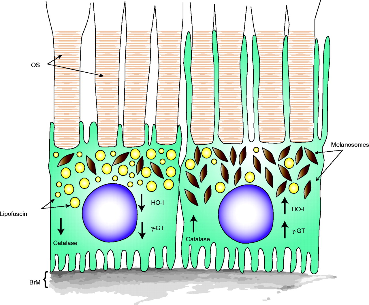
Adjacent RPE cells with differing properties, and their overlying photoreceptors. The left RPE cell might be at greater risk for oxidative damage over time because of its lower abundance of photoprotective melanosomes (brown granules), higher abundance of photoreactive lipofuscin (yellow granules), and lower levels of antioxidant enzymes catalase, heme-oxygenase-1 (HO-1), and γ-glutamyltranspeptidase (γGT). The left cell also has an apical domain of short microvilli interacting with overlying photoreceptors, a property that correlates with the patchy expression of E-cadherin in the RPE (27). The cell on right is distinct in each of these properties. Also illustrated is Bruch’s membrane (BrM) on the basal side of the RPE, and photoreceptor outer segments (OS) embedded in RPE apical microvilli. The microvilli vary in length between the two cells.

