- Institution: Stanford Univ Med Ctr Lane Med Lib/Periodical Dept/Rm L109
- Sign In as Member / Individual
Phosphoinositide Phosphatases: Emerging Roles As Voltage Sensors?
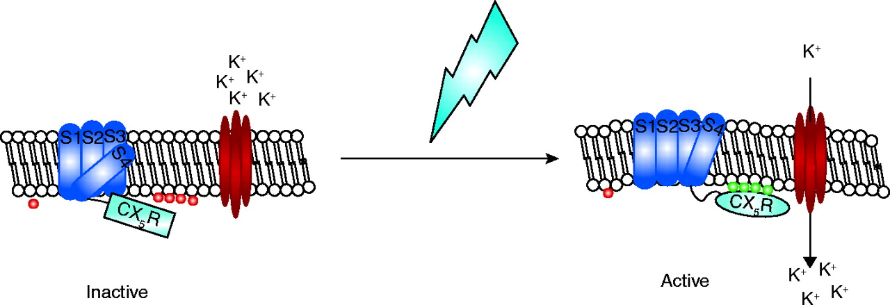
Schematic of potential mechanisms of action of Ci-VSP. The sensor portion of Ci-VSP is shown as dark blue barrels labeled S1–S4 and the phosphatase domain by a light-blue box (inactive) or oval (active). PI(3,4,5)P3 is indicated by red circles whereas PI(4,5)P2 is represented by green circles. The red ovals represent an inwardly rectifying potassium channel (Kir) and the lightning bolt indicates a change in membrane potential and/or conformation. In the absence of a change in membrane shape or potential, Ci-VSP is in its inactive state and the nearby K+ channel is not active. A change in the membrane (lightning bolt) is sensed by the S4 transmembrane domain of Ci-VSP, which then undergoes a conformational change resulting in the activation of the phosphatase. This could occur either by repositioning the phosphatase domain closer to its substrate and/or by changing its conformation thereby activating it. In either case, the localized concentration of PI(4,5)P2 (green circles) in the membrane is increased leading to increased activity of a closely juxtaposed channel.

