- Institution: Stanford Univ Med Ctr Lane Med Lib/Periodical Dept/Rm L109
- Sign In as Member / Individual
SAR by NMR: Putting the Pieces Together
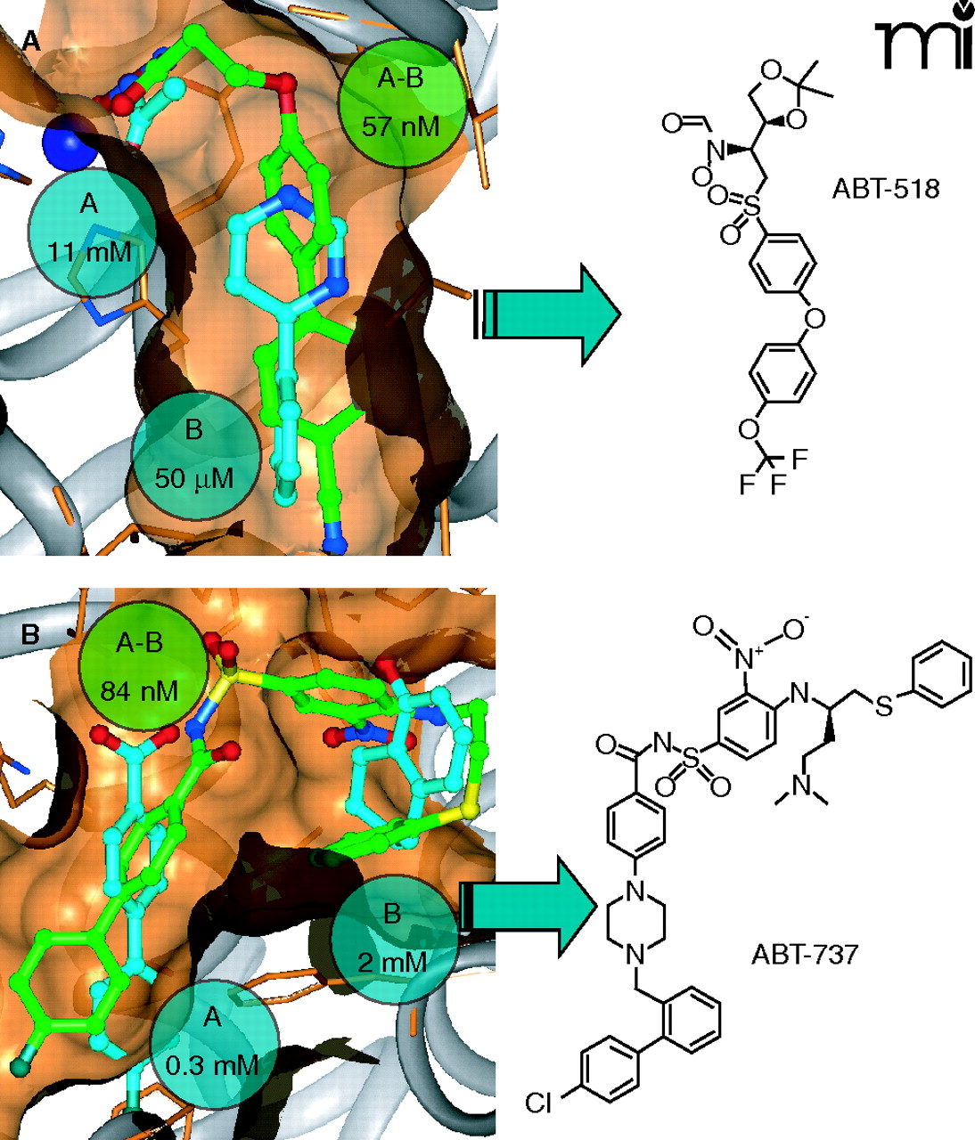
Figure 4.
SAR by NMR on matrix metalloproteinases and Bcl-2 proteins. In each case, matrix metalloproteinases (A) (28–30) and Bcl-2 proteins (B) (31, 32), the identified fragment leads are shown with cyan carbons, whereas the linked compounds are denoted with green carbon atoms. All structures were experimentally determined by NMR. The measured potencies for the individual fragments and the linked compounds are given in cyan and green spheres, respectively. The chemical structure of the final optimized inhibitors (both of which exhibited sub-nM potency in vitro) are shown to the right.

