Bridging Knowledge Gaps on the Long Road to Regeneration: Classical Models Meet Stem Cell Manipulation and Bioengineering
Regeneration research established its roots in the eighteenth century when it firmly debunked the established theories of reproduction by disproving pre-formation. It was then that the classical models (invertebrates and salamanders) exemplified their astonishing capabilities for regenerating body parts. Since that time, regeneration has captured the fascination of scientists and has been often portrayed as one of the most important scientific questions to be answered (1).
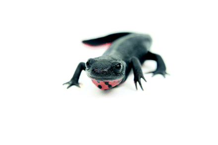
In our time, regeneration research has come into the spotlight through recent molecular exploitation of the classical models as well as through a better (albeit partial) understanding of the potential of somatic and embryonic stem cells. However, no serious research effort has been exerted at the interface between these two areas, which, rather than being disparate and exclusive, should be viewed as complementary. It is well known that newts, for example, regenerate amputated limbs or removed lenses and retina through trans-differentiation of terminally differentiated somatic cells at the site of injury (1). Stem cells, on the other hand, are involved in repair of damaged tissues and do not have to be local—they can be recruited from sites distal to the site of injury (2) and can be stimulated to differentiate by specific damage-indicating signals. Maintenance of their undifferentiated state is controlled by several factors. Different stem cells also possess a molecular signature that characterizes their “stemness” state (i.e., true characteristics of stem cells). On the other hand, somatic newt cells must “dedifferentiate” and lose their tissue characteristics in order to become undifferentiated. Do these dedifferentiated cells of newts show any stemness that could categorize them as stem cells, thus uniting the classical models of repair and regeneration with repair mechanisms in higher mammals? Such a hypothesis was first presented to illustrate possible similarities of limb blastema cells (responsible for newt limb regeneration) and mesenchymal stem cells (3). If the answer to the question is “yes,” then subsequent research might reveal similar strategies for regeneration that have been maintained throughout evolution. Thus, the classical models are imperative partners to stem cells in the quest for repairing and regenerating damaged or lost tissues and should not be neglected. A few recent papers on these topics emphasize this kind of relationship (4–6). Mouse fibroblasts can be reverted to pluripotent stem cells by retroviral introduction of Oct3/4, Sox-2, c-Myc and Klf4. Oct3/4 and Sox-2 are considered to be stem cell pluripotency-maintaining factors (Box 1). These pluripotent stem cells are similar to embryonic stem cells, but they still differ when it comes to gene expression and methylation and also fail to produce adult chimaeras. This means that when such cells were implanted into the blastocyst they did not contribute to tissue production. Such a test is performed to verify that these cells are really multipotent stem cells and able to maintain their biological activity. However, when these cells are selected for Nanog (another pluripotency-maintaining factor) production, they become germline-competent. This means that when a chimeara was crossed with a normal mouse it led to birth of offsprings with the genetic content of the induced pluripotent stem cells and show ES cell-like gene expression and methylation (4, 5). These studies clearly indicate that differentiated cells can be reprogrammed to achieve a stem cell–like state. In this sense, these reprogrammed cells behave like the reprogrammed cells during limb regeneration in the newt. However, in newt limb regeneration and during dedifferentiation, the reprogrammed cells do not really become bona fide embryonic stem cells. They become only multipotent progenitor cells to assure correct regeneration. To what degree the newt cells are capable of returning to pluripotent stem cells in unknown. One of the first attempts to illustrate this was recently reported by examining the expression of nucleostemin, a stem cell– and cancer cell–specific protein. During lens regeneration, nucleostemin accumulates in the dedifferentiated pigment epithelial cells that are responsible for regenerating the lens, indicating that differentiated newt cells do undergo changes that make them more similar to stem cells (6).
Totipotent, Pluripotent, & Multipotent Cells
Totipotent stem cells: Cells able to give rise to all embryonic somatic cells and germ cells. In other words, they can build a whole animal. The zygote and a few early cells of the morula are totipotent.
Pluripotent stem cells: These cells are descendants of totipotent stem cells and can give rise to cells of the three germ layers: endoderm, mesoderm, and ectoderm. They have no contribution to extraembryonic membranes or the placenta.
Multipotent stem cells: These produce cells of a particular lineage or closely related family.
All these issues bring up the next question: How will regeneration research make an impact in the biomedical field? There are two major challenges of regeneration research. One is to gain understanding of the extraordinary biological phenomenon and explain its relationship to development and evolution. Because a trait should be beneficial to the organism, regeneration (for example, in newts) should make sense in terms of evolutionary success. Nevertheless, at this point, it is quite difficult to answer why regenerating a lens is advantageous to a newt and not to a primate. Therefore, the biology of the newt might be unique for regeneration. And because of this, gene regulation could be governed by unsuspected mechanisms. Such mechanisms could also be shared by stem cells as well, under the umbrella of the stemness mentioned above. The second challenge is to use this knowledge to find cures for replacing damaged tissues. The need for organs to be transplanted is great but the supplies are limited. Contrary to the popularization of the issue, however, answering the questions of one challenge does not necessarily mean that the other will be achieved. In other words, even if we understand how the newt regenerates a limb, it does not necessarily follow that we would accomplish the same feat in humans. The reason might have to do with unique animal biology or other evolutionary constraints. It would be of great benefit if salamanders could teach us how to grow lost legs and repair damaged brains, but how realistic is such an expectation? If, in the end, we do face such a deterrent, what is the future of regeneration?
The solution might be provided from another research front. Recent advances in bioengineering are quite promising for building-up tissues. Scaffolds, local cells, and growth factors have been utilized to engineer tissues (7, see also 8). Research on biomaterials—especially for 3-D scaffolds—is important because factors such as matrix stiffness can direct stem cell differentiation (9, 10); however, although this finding might be applicable to the formation of tissues (e.g., skin, bladder, or bone) it is highly unlikely that tissue engineering will replace an amputated leg with a replica of it, as is seen in the newt. Successful reconstruction of damaged organs stands a much better chance if we combine knowledge from both the spheres of biology and engineering. For example, basic biology experiments with the classical models (transdifferentiation) or with stem cells can reveal how to differentiate lens cells in vitro (11, 12). The use of appropriate scaffolds then could lead to the build-up of a lens of desired size and shape to be implanted in vivo to replace a cataractous lens. A lost finger can be reconstructed by producing the different tissues separately and piecing them together. To achieve successful transplantation of autograft organs (i.e., “self donation” or the growth of tissues and organs from cells that originate from the affected organism) organs or organ regeneration in situ the best design for proper regeneration will make use of all possible avenues. Thus, “directed regeneration” should span the principles of classical regeneration models, stem cell differentiation, and tissue engineering to make the long road to regeneration a smooth journey.
- © American Society for Pharmacology and Experimental Theraputics 2007
References

Panagiotis A. Tsonis, PhD, is a Professor of Molecular Biology and the Director of the Center for Tissue Regeneration and Engineering at the University of Dayton, Ohio. He is very interested in many issues of tissue regeneration, the mechanisms of transdifferentiation, and induction of regeneration in mammals. E-mail Panagiotis.Tsonis{at}notes.udayton.edu; fax 937–229–2021.


