- Institution: Stanford Univ Med Ctr Lane Med Lib/Periodical Dept/Rm L109
- Sign In as Member / Individual
G Protein-Coupled Receptors Go Extracellular: RhoA Integrates the Integrins
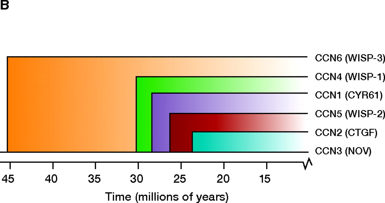
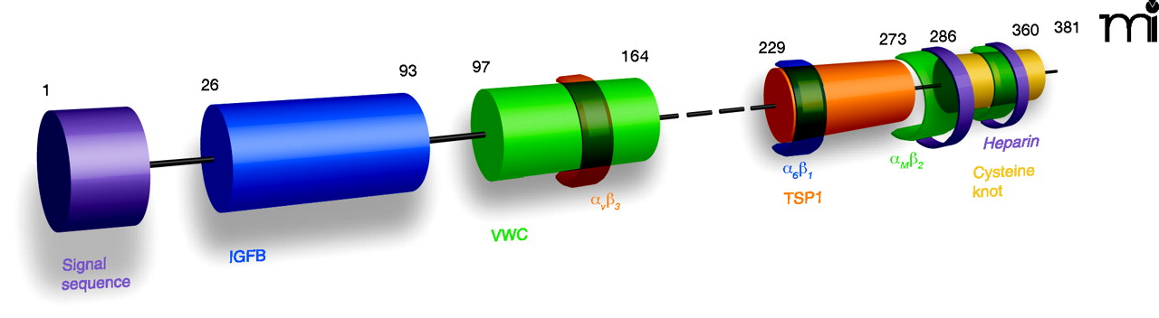
The CCN family of proteins. A modular representation of the human CCN1 protein indicates i) the signal sequence; ii) the insulin-like growth factor–binding protein homology domain (IGFB, blue); iii) the von Willebrand factor type C domain (VWC, green); iv) the thrombospondin type 1 homology domain (TSP1, orange); and the cysteine knot–containing C-terminal domain (yellow). Semi-transparent colored bands around the domains indicate sites that interact with integrins (e.g., integrin αvβ3, α6β1, and αMβ2); also shown are the two binding sites for heparin (purple C-terminal bands). The dashed line indicates the hinge region; numbers represent amino acid positions in the protein sequence. The dendrogram reflects sequence conservation among the human CCN proteins (20). See text for details.

