- Institution: Stanford Univ Med Ctr Lane Med Lib/Periodical Dept/Rm L109
- Sign In as Member / Individual
Inside Information: The Unique Features of Visceral Sensation
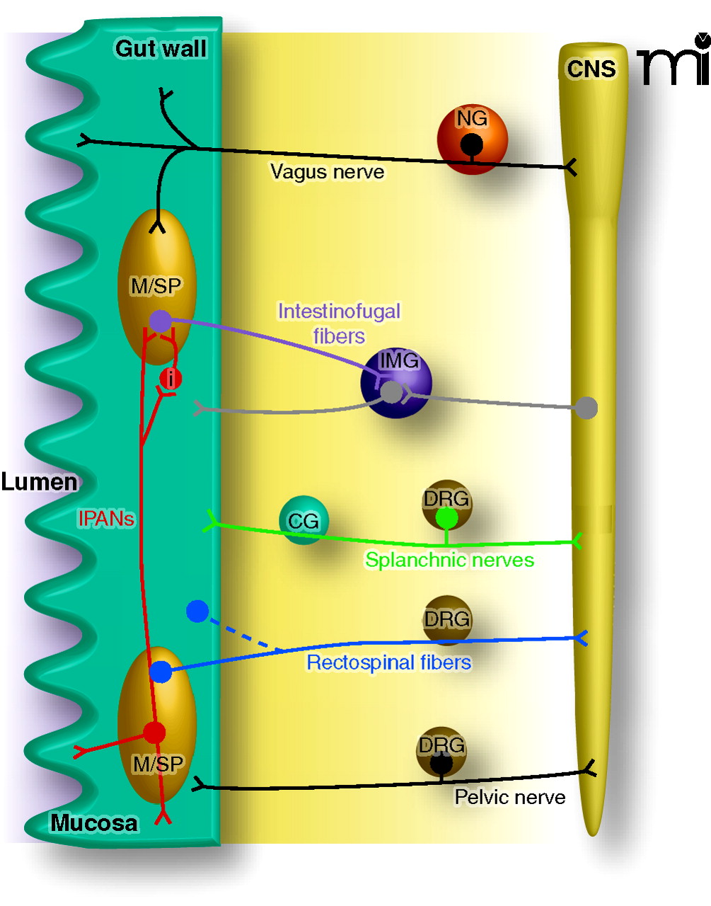
Functional neuroanatomy of the visceral sensory system. The gut is depicted here as an example of the sensory innervation of the visceral system. Vagal afferent neurons, which do not innervate the urinary bladder or the distal gut, have cell bodies in the nodose ganglia (NG) bilaterally and travel alongside parasympathetic efferent pathways to organs in the thoracic and abdominal cavities. Once in the gut wall, vagal afferent fibers innervate neurons in the myenteric or submucosal plexus (M/SP), circular and longitudinal muscle layers, and the mucosa. Pelvic afferent neurons also travel alongside parasympathetic efferent pathways, but their cell bodies are in dorsal root ganglia (DRG). Other spinal nerves (e.g., greater splanchnic) travel alongside sympathetic efferent pathways, have cell bodies in DRG, and pass through prevertebral ganglia (e.g., the celiac ganglion, CG). Intestinofugal afferents (purple) synapse onto efferent sympathetic neurons in prevertebral ganglia, such as the inferior mesenteric ganglion (IMG) and have their cell bodies in M/SP. Afferent fibers of the intrinsic (or enteric) nervous system, termed intrinsic primary afferent neurons (IPANs), synapse onto intestinofugal fibers, either directly, or via interneurons (i). Rectospinal fibers (blue) have cell bodies in the myenteric plexus or muscle layers, with axons terminating in the spinal cord (CNS). Note that not all these nerves and fibers will terminate in the same areas of the gut, and inputs to the spinal cord may traverse a number of different levels; this figure has been simplified for clarity.

