Fragment-based ligand discovery
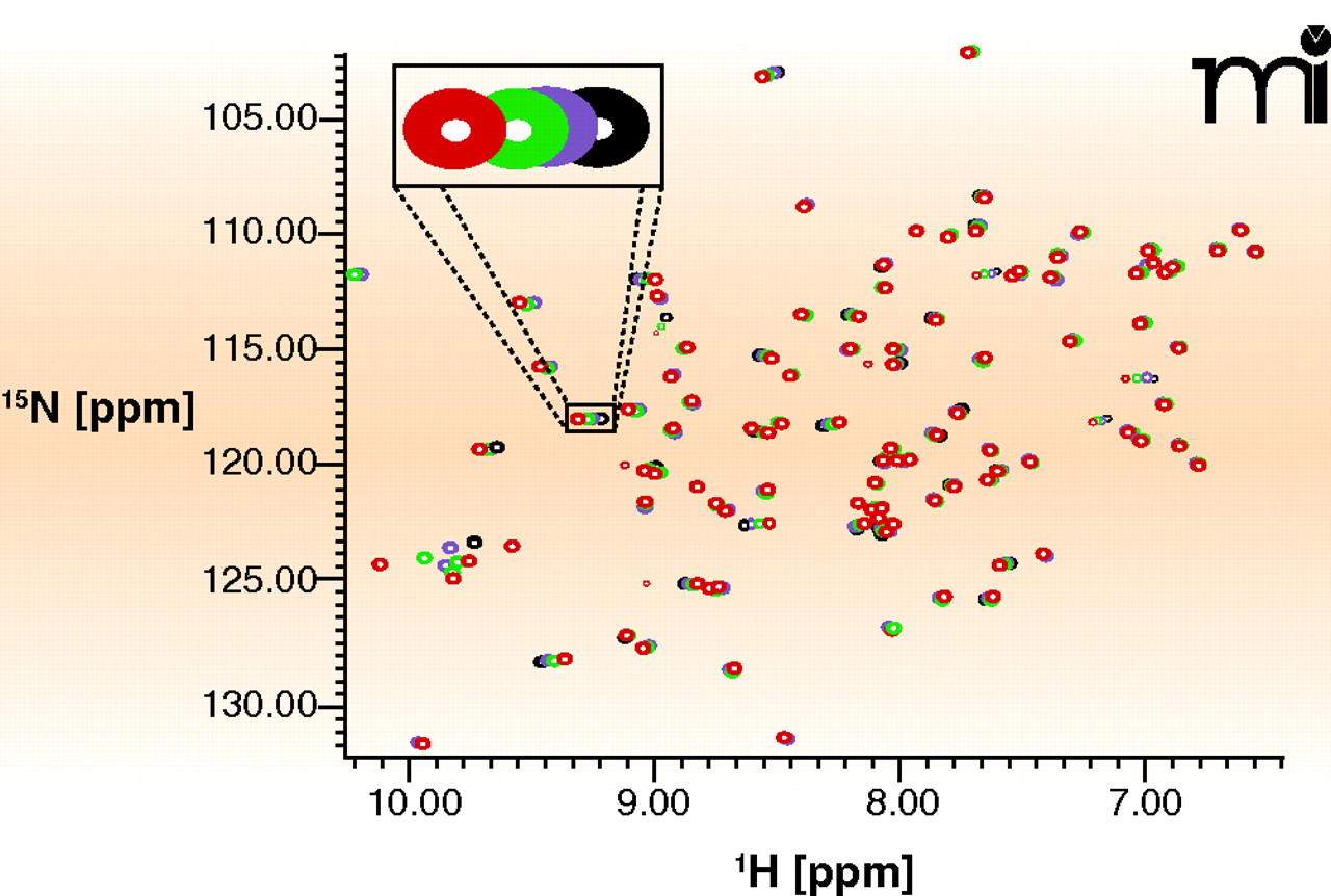
Figure 4.
15N NMR—HSQC measurements. Each peak in the NMR experiment reports on the local chemical environment of a nitrogen atom; this environment changes during compound binding and is reported as shifts in peaks. The method requires large quantities of 15N-labeled protein that must be stable at 100 μM in low-salt buffer for many days. Here, a representative HSQC spectrum of the protein Pin1 with detail showing how peak positions shift (schematized by changes in color of peak) as concentration of a ligand is increased (J. Murray, Vernalis, unpublished results).

