- Institution: Stanford Univ Med Ctr Lane Med Lib/Periodical Dept/Rm L109
- Sign In as Member / Individual
Pharmacokinetic Assessment of Multiple ATP-binding Cassette Transporters: The Power of Combination Knockout Mice
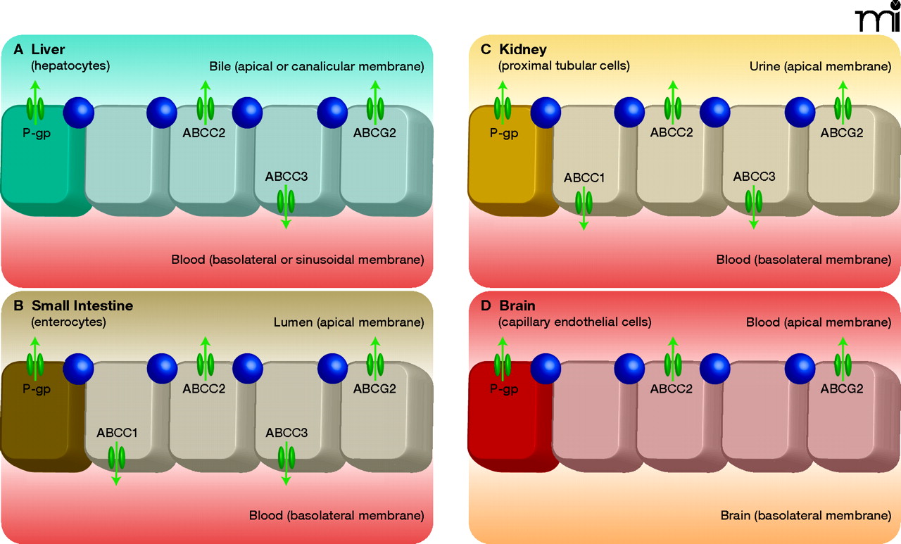
Tissue distribution of the human ATP-binding cassette (ABC) transporters ABCB1 (P-gp), ABCG2, and ABCC1-3. A) In the liver, P-gp, ABCG2, and ABCC2 are located in the canalicular (apical) membrane of hepatocytes, pumping their substrates into bile. ABCC3 is present in the sinusoidal (basolateral) membrane of hepatocytes, and pumps its substrates towards the blood circulation. Notably, it was recently shown that Abcc1 is present in activated rat hepatic stellate cells (HSCs), but not in hepatocytes (64). B) In the small intestine, P-gp, ABCG2, and ABCC2 in the luminal (apical) membrane of enterocytes pump their substrates into the intestinal lumen. ABCC1 and ABCC3, located in the basolateral membrane of enterocytes pump their substrates towards the blood circulation. C) In the kidney, P-gp, ABCG2, and ABCC2 are localized in the apical membrane of proximal tubular cells and extrude their substrates into the urine. ABCC1 and ABCC3 are present at the basolateral membrane, pumping their substrates towards the blood circulation. D) At the blood-brain barrier, P-gp, ABCG2, and ABCC2 are located at the apical membrane of capillary endothelial cells, pumping their substrates towards the blood. [ABCC1 (not shown) is also expressed in the basolateral membrane of epithelial cells of the choroid plexus, preventing the entry of its (potentially harmful) substrates into the cerebrospinal fluid (CSF); see (1).] (Tight junctions are indicated by circles.)

