- Institution: Stanford Univ Med Ctr Lane Med Lib/Periodical Dept/Rm L109
- Sign In as Member / Individual
Chemokine Signaling and the Management of Neuropathic Pain
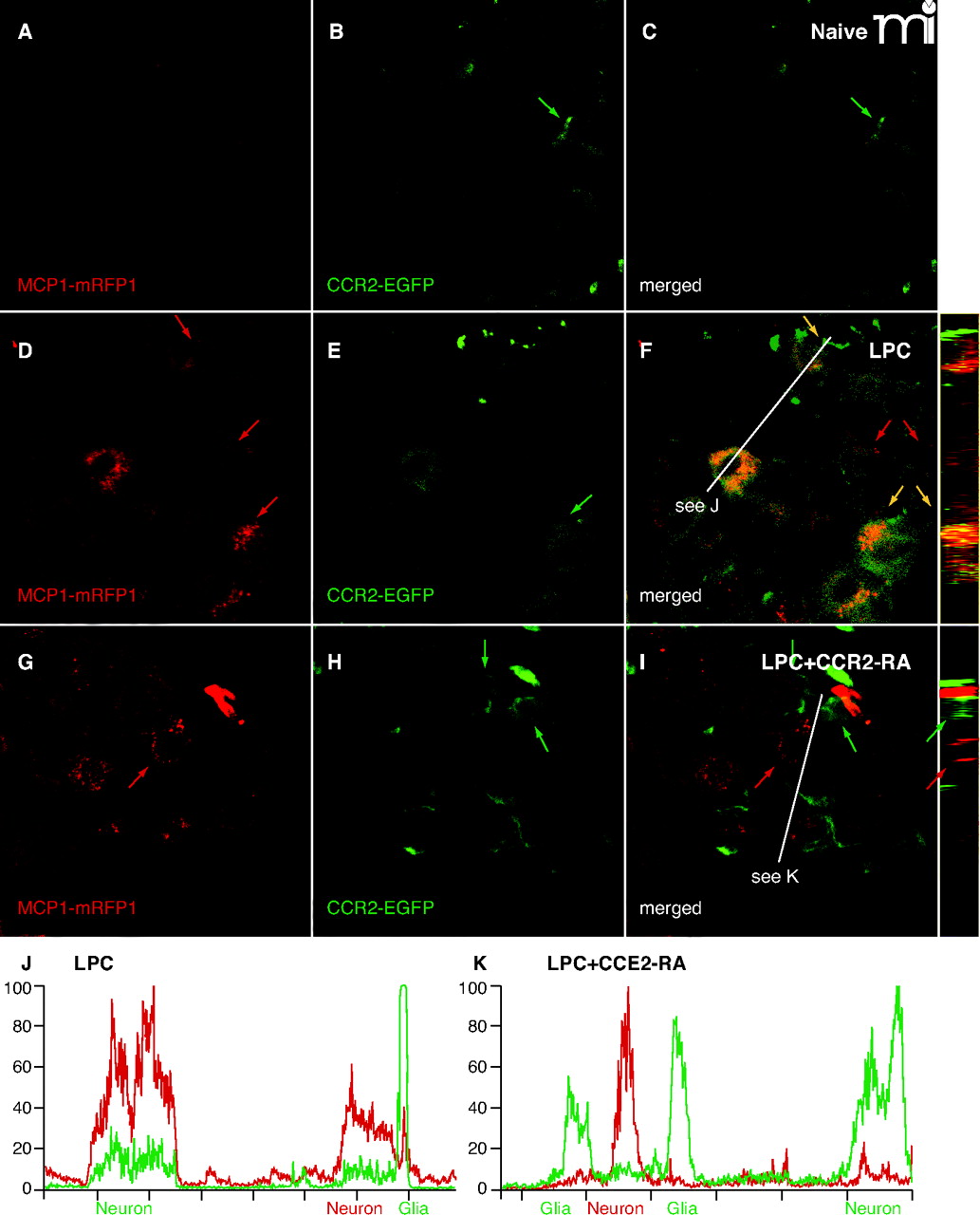
CCR2 signaling is activated in the DRG. Mice expressing MCP1 tagged with monomeric red fluorescent protein-1 (MCP1–mRFP1) were crossed with mice expressing CCR2 tagged with extra green fluorescent protein (CCR2–EGFP) mice. The resultant transgenic mice were subjected to lysophosphatidylcholine-(LPC)-induced demyelination of the sciatic nerve. In the DRG ipsilateral to the injury, expression of both MCP1 and CCR2 increased (D–F), whereas there was little expression of MCP1 or CCR2 under naive conditions (A–C). MCP1–mRFP1 mainly localized to neurons (large red arrow) and, to some extent, to satellite glia [small red arrow (D)]. CCR2–EGFP localized to neurons (large green arrow) and satellite glia [small green arrow (E)]. Most CCR2–EGFP-expressing neurons and satellite glia also contained MCP1–mRFP1 [yellow arrow (F)]. Injection of the CCR2–RA eliminated MCP1–mRFP1 in satellite glia [small green arrow (G–I)]. Also, after CCR2–RA treatment, MCP1–mRFP1- and CCR2–EGFP-expressing cells existed as separate populations [large green and red arrows(G–I)]. Cross-sectional images across the white lines are shown right to (F) and (I). (J, K) Intensities of mRFP1 and EGFP in shorter white lines in (F) and (I) are expressed in arbitrary units to compare relative signal intensities among different cells. (J) In the LPC group, most neurons that express CCR2–EGFP also contain MCP1–mRFP1. Also, the CCR2–EGFP signal in neurons is relatively weaker than the signal in satellite glia (J). (K) In the LPC plus CCR2–RA group, however, most CCR2–EGFP-expressing cells do not contain significant amount of MCP1–mRFP1 signal. In addition, the CCR2–EGFP signal in neurons is now as strong as the signal in satellite glia (K).

