- Institution: Stanford Univ Med Ctr Lane Med Lib/Periodical Dept/Rm L109
- Sign In as Member / Individual
Tumor-associated, Estrogen Receptor-related Antigen EBAG9: Linking Intracellular Vesicle Trafficking, Immune Homeostasis, and Malignancy
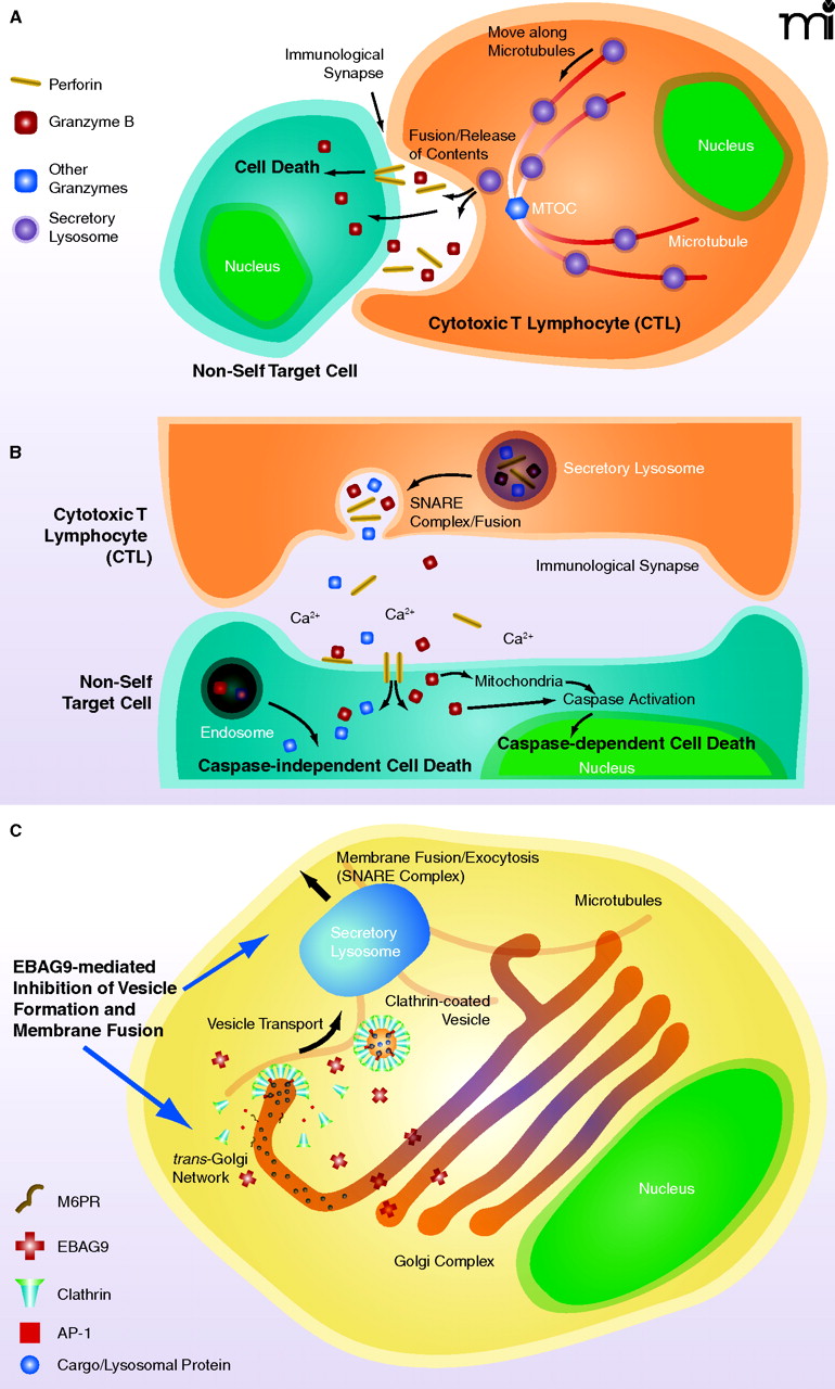
Formation and secretion of secretory lysosomes in the cytotoxic T-lymphocyte (CTL). A. Release of secretory lysosomes by CTLs when these cells encounter, recognize and interact with non-self cells. CTLs recognize non-self cells through foreign antigens co-presented with NHC class I molecules in the surface of target cells, forming with them an “immunological synapse.” Intra-CTL secretory lysosomes associate with microtubules and translocate towards the microtubule-organizing center (MTOC), accumulating at the vicinity of immunological synapses. Accumulated secretory lysosomes then fuse to the CTL plasma membrane and secrete their contents into the immunological synapses through the SNARE complex. Released perforin molecules allow entry of granzymes into the cytoplasm of the target cells, activating apoptotic pathways and leading to their demise. B. Secretory lysosome-mediated cell death by CTLs. Upon activation of CTLs, and after formation of the immunological synapse with non-self target cells, CTLs release the contents of secretory lysosomes (perforins and granzymes) into the lumen of the immunological synapse through reverse pinocytosis organized by the SNARE complex. Perforin then forms a complement-like transmembrane pore in the plasma membrane of target cells under the influence of Ca2+, allowing transfer of granzymes into these cells, in addition to uptake of granzymes by pinocytosis. Intracellularly accumulated granzymes stimulate cas-pase-dependent and -independent pathways to finally kill target cells (4). C. Formation and maturation of secretory lysosomes in CTLs. Lysosomal proteins, such as perforin and granzymes, are produced in the endoplasmic reticulum and transferred to the Golgi complex. These molecules then accumulate in the trans-Golgi network (TGN), where they interact with sorting molecules, the mannose 6-phosphatase receptors (M6PRs). TGN membranes containing lysosomal proteins eventually form clathrin-coated vesicles by attracting clathrins and adaptor proteins (APs) (e.g. AP-1). Clathrin-coated vesicles move along microtubules and fuse to maturing secretory lysosomes.

