- Institution: Stanford Univ Med Ctr Lane Med Lib/Periodical Dept/Rm L109
- Sign In as Member / Individual
Translational NeuroImaging of the CNS: Novel Pathways to Drug Development
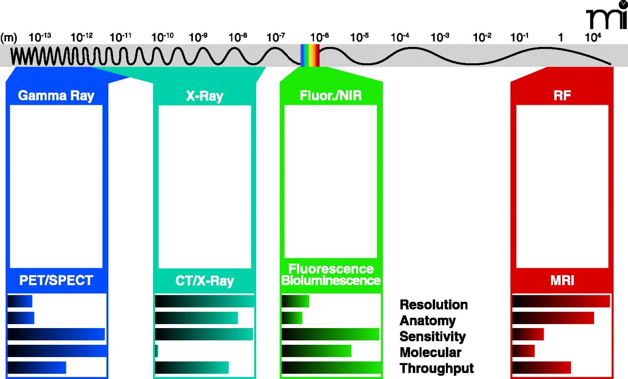
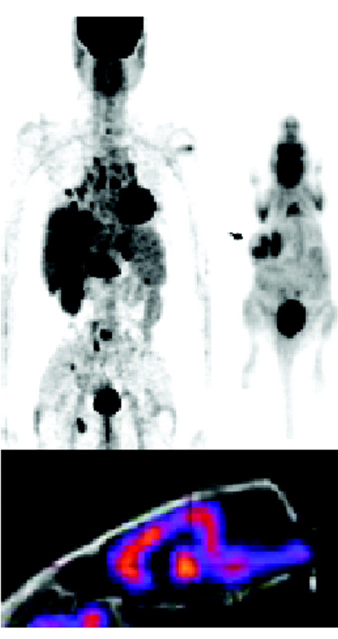

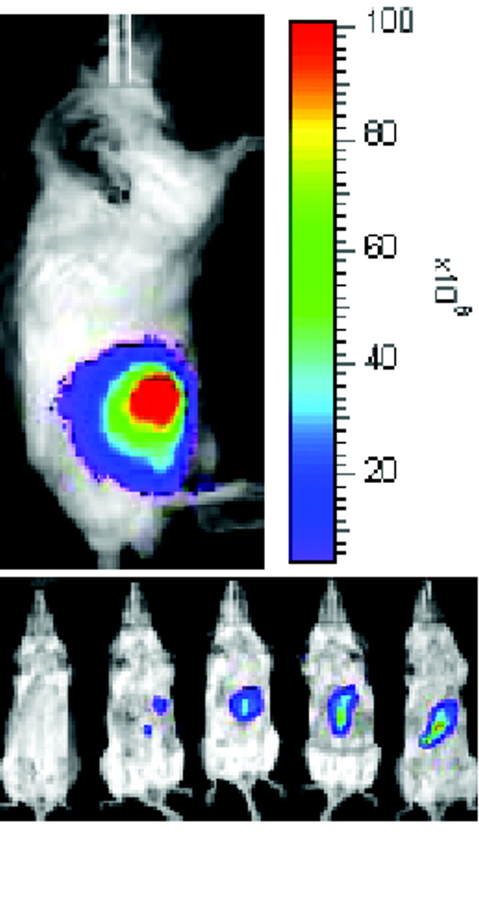
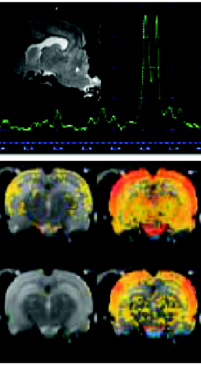
The spectrum of neuroimaging techniques: Specialization and complementarity. All imaging technologies utilize specific electromagnetic wavelengths to generate and measure signals. MRI utilizes radio frequencies at the far right of the electromagnetic spectrum and maintains a high static magnetic field to produce images of high spatial resolution. MRI provides anatomical and some physiological imaging. In contrast, fluorescence/bioluminescence and near-infrared optical imaging utilize visible or near-visible wavelengths to produce images of high sensitivity relatively quickly, but the spatial resolution is lower than that provided by MRI. In vivo optical imaging can provide additional information at the molecular level. More energetic and shorter wavelengths are employed in X-ray and CT scanning, which provide excellent anatomical images of high sensitivity but little information at the molecular level. PET and SPECT imaging, which utilize very short wavelengths—in the gamma range of the spectrum—provide excellent sensitivity and yield information about metabolism, molecular targets, and physiology, although spatial resolution may be better provided by other modalities. (See text for discussion of methods and modalities for translation into human studies and therapeutics.)

