
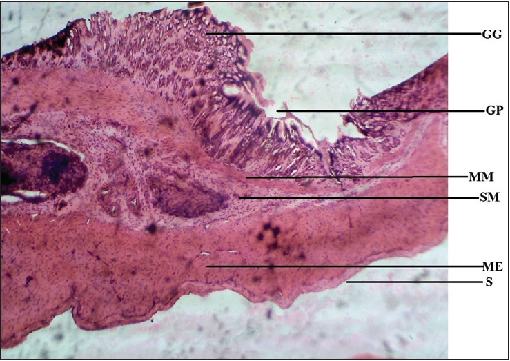
Figure 6: Photomicrograph of cane toad stomach showing gastric gland (GG), gastric pit (GP), muscularis mucosae (MM), submucosa (SM), muscularis externa (ME), and serosa (S). Stained with H&E (40×)
| Close | |
|
|
|
|
Figure 6: Photomicrograph of cane toad stomach showing gastric gland (GG), gastric pit (GP), muscularis mucosae (MM), submucosa (SM), muscularis externa (ME), and serosa (S). Stained with H&E (40×)
|
|