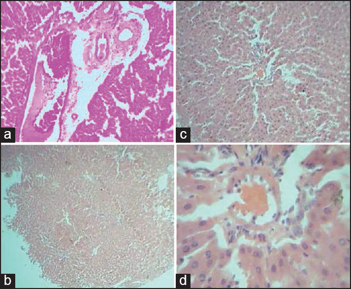
Figure 1: Comparative histology illustrations of the mammalian liver. (a) Section of the Liver of rattus norvergicus showing hepatocytes (he), and portal triad (pt) H&E × 100 (b) Section of the Liver of rattus norvergicus Gallus gallus domesticus domesticus agama aculeata sp rana tigrinas clarias gariepinus showing hepatocytes (he) disposed in cord and sheet H&E × 40 (c) Section of the Liver of rattus norvergicus showing hepatocytes (he) disposed in cord and sheet. A prominent central venules (cv) is present in the center H&E × 100 (d) Section of the liver of rattus norvergicus showing hepatocytes (he) disposed in cord and sheet. A prominent central venules (cv) is present in the center H&E × 400