Close
  Indian J Med Microbiol
 

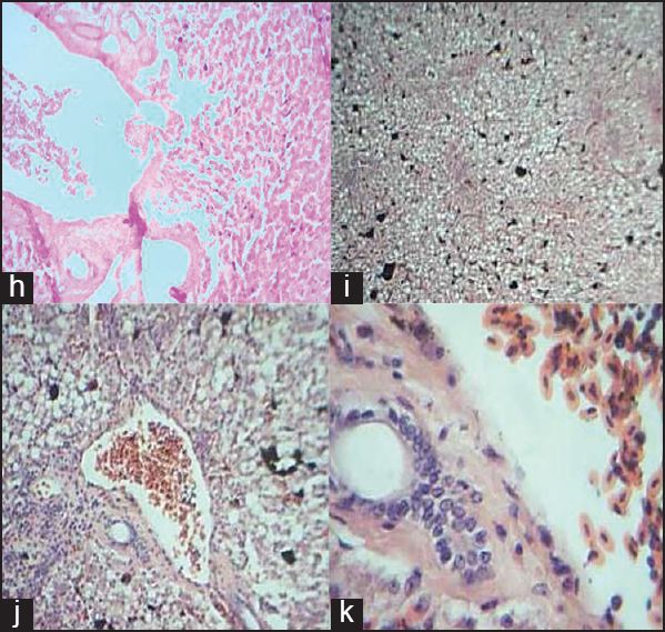
Figure 3: Comparative histology illustrations of the reptile liver. (h) Section of the liver of Agama aculeata sp showing hepatocytes (he) in singles and little clusters H&E × 40 (i) Section of the liver of Agama aculeata sp showing hepatocytes disposed in singles and little clusters separated by thin fi brovascular stroma. Portal vein, bile ductile and dark staining artefacts are present H&E × 100 (j) Section of the liver of Agama aculeata sp showing hepatocytes disposed in singles and little clusters separated by thin fibrovascular strom, dark staining artefacts are present H&E × 40 (k) Section of the liver of Agama aculeata sp showing portal vein and bile ductile (bd) with nucleated red blood cells (rb) H&E × 100

Figure 3: Comparative histology illustrations of the reptile liver. (h) Section of the liver of <i>Agama aculeata sp</i> showing hepatocytes (he) in singles and little clusters H&E × 40 (i) Section of the liver of <i>Agama aculeata sp</i> showing hepatocytes disposed in singles and little clusters separated by thin fi brovascular stroma. Portal vein, bile ductile and dark staining artefacts are present H&E × 100 (j) Section of the liver of <i>Agama aculeata sp</i> showing hepatocytes disposed in singles and little clusters separated by thin fibrovascular strom, dark staining artefacts are present H&E × 40 (k) Section of the liver of <i>Agama aculeata sp</i> showing portal vein and bile ductile (bd) with nucleated red blood cells (rb) H&E × 100