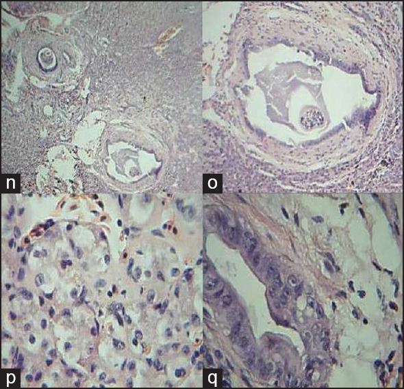
Figure 5: Comparative histology illustrations of the fish liver. (n) Section of the liver of Clarias gariepinus showing sheets and clusters of hepatocytes H&E × 40 (o) Section of the liver of Clarias gariepinus showing a biliary duct within the liver parenchyma with kuffer cell (kc), H&E × 100 (p) Section of the liver of Clarias gariepinus showing hepatocytes disposed in clusters (he). Few nucleated red cells (rc) are seen in the background H&E × 400 (q) Section of the liver of Clarias gariepinus showing a biliary duct (bd) with straitified columnar epithelium. Few hepatocytes and nucleated red cell are seen in the periphery (H&E × 400)