
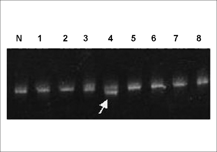
Figure 1: Part of CSGE gel showing the migration pattern of exon 14D amplified fragments. Fragments with abnormal migration patterns are marked by arrow. N: normal control and 1, 2, 3, 4 (HA11), 5, 6, 7, 8, 9 different hemophilia A patients.
|
|
Close |
|
Figure 1: Part of CSGE gel showing the migration pattern of exon 14D amplified fragments. Fragments with abnormal migration patterns are marked by arrow. N: normal control and 1, 2, 3, 4 (HA11), 5, 6, 7, 8, 9 different hemophilia A patients.
|
|