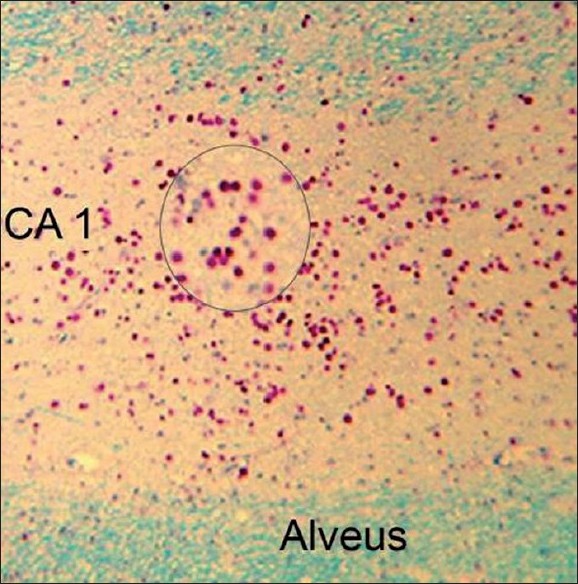
Figure 1: Microphotograph showing dense deposition of corpora amylacea (CoA) in the CA1 sector of the hippocampus in a 42-year-old male with mesial temporal lobe epilepsy and hippocampal sclerosis. Inset shows a magnifi ed view of the CoA (Luxol-fast blue– Periodic Acid Schiff stain, X150). 83 mm x 84 mm (300 x 300 DPI)