
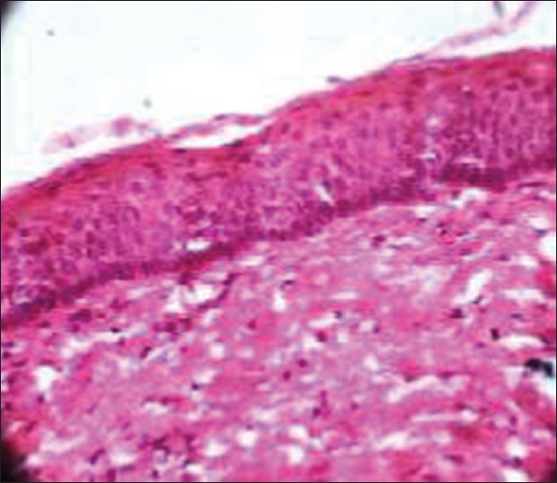
Figure 4: Histological section showing stratified squamous parakeratinized epithelium with palisading pattern of columnar cells along with keratin flakes suggestive of odontogenic keratocyst under high power (×40)
|
|
Close |
|
Figure 4: Histological section showing stratified squamous parakeratinized epithelium with palisading pattern of columnar cells along with keratin flakes suggestive of odontogenic keratocyst under high power (×40)
|
|