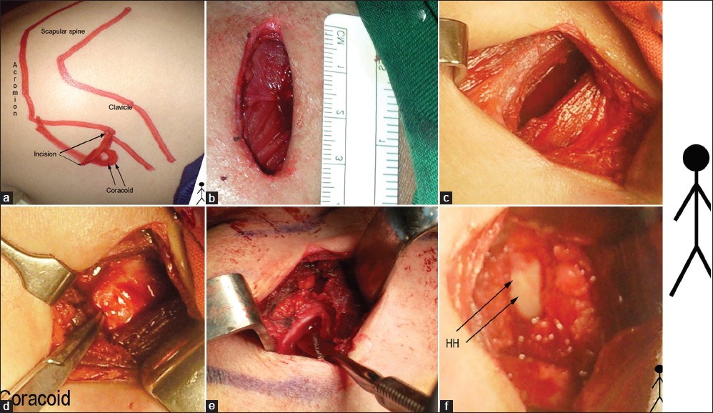
Figure 1: Intraoperative photos of the surgical technique (This is the Right shoulder, head, and feet of the patient indicated by the stick figure to the right of the figure) (a) Outline of the bony landmarks and the incision. (b) The 3 cm incision. (c) The deltopectoral interval. (d) The coracoid. (e) Dissector under the CHL (f) Capsulotomy performed and the Humeral Head (HH) shown