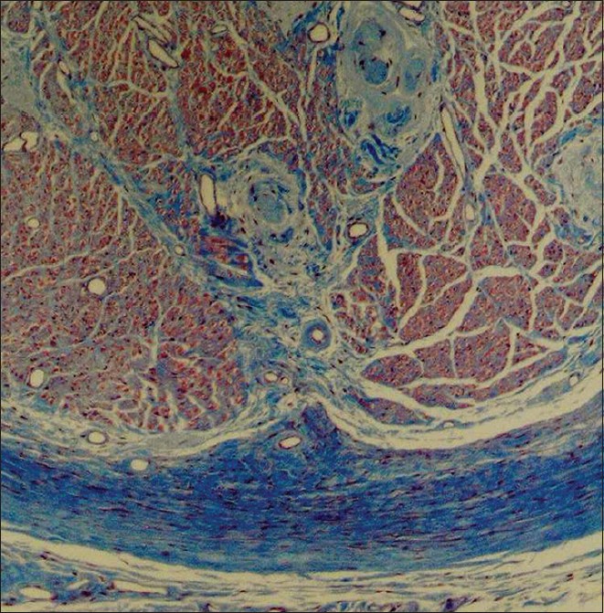
Figure 3: Histologic specimen noting signs of suprascapular nerve compression seen in cadavers with an ossified suprascapular ligament. This section is from the suprascapular nerve distal to the ossified ligament and notes vascular hyalinization and thickening of the epi and perineuria (Trichrome ×44)