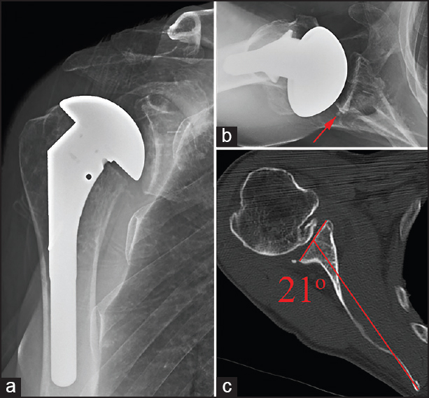
Figure 3: Grade C seating - Immediate postoperative anteroposterior radiograph (a), axillary radiograph (b), and preoperative axial computed tomography scan with measured retroversion (c) illustrating an example of Grade C glenoid seating following total shoulder arthroplasty. Preoperative retroversion was incompletely corrected, leaving an uncontained area of the posterior glenoid which was filled with a wedge of cement (red arrow) encompassing just under 50% of the glenoid surface seen only on the axillary radiograph