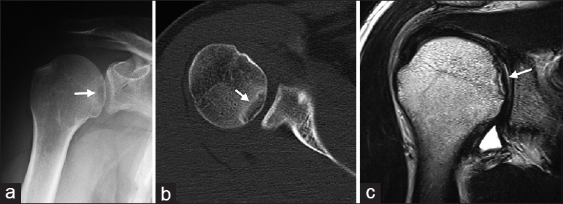
Figure 1: Preoperative imaging findings. (a) Antero-posterior plain radiogram shows the radiolucent lesion in the medial aspect of the right humeral head (white arrow). (b) Axial computed tomography scans show the 2-cm radiolucent lesion surrounded by osteosclerotic change, whereas the subchondral bone maintains its continuity above the lesion (white arrow). (c) Magnetic resonance imaging shows an osteochondral fragment, which is separated from the humeral head, on T2-weighted images. The articular cartilage maintains its continuity (white arrow)