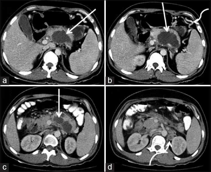
Figure 1: Axial section computed tomography demonstrates nonenhancing cystic lesion in the pancreas with ill-defined margins (straight white arrow a, b, and c). Enlarged lymph nodes in the left para-aortic region with central necrotic component (curved white arrow in d). Collateral vessels (curved white arrow in b) secondary to narrowing of splenic vein