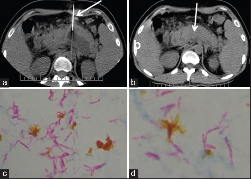
Figure 2: Computed tomography FNA performed with spinal puncture needle (straight white arrow a). After computed tomography guided aspiration drainage, near complete resolution of the collection, was confirmed (straight arrow in b). Microscopy of pus aspirate in Ziehl-Neelsen Stain demonstrates numerous acid fact bacilli (c and d)