|
 
 |
| ORIGINAL ARTICLE |
|
| Year : 2014 | Volume
: 9
| Issue : 4 | Page : 147-150 |
|
Histopathological pattern of cervical cancer in Benin City, Nigeria
Chukwuemeka Asouzu Okoye
Department of Pathology, Federal Medical Centre, Asaba, Delta State, Nigeria
| Date of Web Publication | 14-May-2015 |
Correspondence Address:
Dr. Chukwuemeka Asouzu Okoye
Department of Pathology, Federal Medical Centre, Asaba, Delta State
Nigeria
 Source of Support: None, Conflict of Interest: None  | Check |
DOI: 10.4103/9783-1230.157057

Background: Cervical cancer is the second most frequent malignancy and a preventable cause of mortality and morbidity in females. Objective: The objective was to describe the relative frequency, pattern, and histological types of cervical cancer in a teaching hospital in South-South Nigeria. Materials and Methods: All histologically diagnosed cases of cervical cancer seen over a 10-year period in the Department of Pathology, University of Benin Teaching Hospital, Benin City, Nigeria were reviewed to determine their histopathological patterns. Results: Four hundred and six cases of cervical cancer accounting for 30.3% of cancers in females and 62.9% of female genital tract malignancies respectively were seen during the 10-year study period. The ages of patients with cervical cancer which ranged between 18 and 99 years with a mean of 51.5 years (standard deviation = 12.8), with most frequent occurrence in 50-59 years age group. Squamous cell carcinoma was the most predominant subtype comprising 84.2% of cases while adenocarcinoma constituted 11.8%. Adenoid cystic carcinoma, adenosquamous carcinoma and metastatic carcinoma accounted for 2.0%, 0.8%, and 1.2% of cases respectively. Conclusion: The relative frequency of cancer of the cervix is high in Benin City, and this should necessitate attention to effective cervical cancer screening to increase detection of preinvasive lesions which in turn will decrease the frequency of cervical cancer. Keywords: Cervical cancer, Female genital tract cancer
How to cite this article:
Okoye CA. Histopathological pattern of cervical cancer in Benin City, Nigeria. J Med Investig Pract 2014;9:147-50 |
| Introduction | |  |
Cervical cancer is a major health problem in the developing world. According to a 40-year review by the World Health Organization (WHO) and International Union against Cancer, the most common cancers in females, world over from 1960 to 69 were those from the cervix, breast, and non-Hodgkin lymphoma; and in 1998, was overtaken by breast cancer. However, the same report documents that in 2002 cervical cancer once again became the most common malignancy in females followed by breast cancer and Kaposi sarcoma in sub-Saharan Africa. [1] At present, cervical cancer is documented as the second most common cancer among females in the world and the most common female genital tract (FGT) cancer. [2],[3] Cervical malignancies also constitute a significant number of surgical pathology reports. Okobia and Aligbe in Benin, Nigeria [4] reviewed 2258 cases of malignancies occurring in both males and females in a 20-year period and observed a predominance of cancers in females (64%); with breast and cervical cancer being the first and second most common cancers, respectively, for women and both accounting for 59% of all cancers in females. While cervical cancer continues to be a significant cause of mortality and morbidity in the developing world, much of the developed world has shown remarkable reduction in cervical cancer deaths due to cervical cancer screening. [5],[6],[7]
The relative frequencies of cervical cancer vary in different regions of the world ranging from the high frequencies obtained in Nigeria and most Sub-Saharan states, and diminishing frequencies found across Asia and the Western world. Similarly, there has been a slight variation in the histological subtypes seen in different areas under review.
| Materials and Methods | | |
All data were obtained from the Department of Pathology, University of Benin Teaching Hospital (UBTH), which receives samples for histology from the clinical departments in the same hospital and three other government-owned hospitals which have no functional pathology departments, as well as from private clinics within Benin Metropolis, neighboring towns and states. Surgical biopsies from FGT malignancies received and reported at the Department of Pathology, UBTH over the 10-year period between January 2000 and December 2009 were used for this study. Demographic data for this study were obtained from surgical pathology registers, duplicate copies of histological histology reports and request cards of all patients with FGT malignancies diagnosed within the study period. Where the age was not stated, corresponding case notes were retrieved from the medical records department. The corresponding slides were retrieved and reviewed by a pathologist and resident and diagnoses re-confirmed. Where the slides were missing or sections not properly preserved, paraffin-embedded tissue blocks were retrieved from the archives of the Department of Pathology, UBTH and new sections were cut and stained with hematoxylin and eosin. In cases where a definitive diagnosis could not be reached, available special stains were used to define the diagnosis. A thorough assessment of names and associated hospital reference numbers of all resection and biopsy specimens were done to prevent double counting of cases. All histologically diagnosed cases of malignancy within the period were also noted so as to calculate the relative frequency of FGT malignancies within the same period. The 2003 WHO histological index of cervical malignancies was used to classify the tumors. [8] All cases without such diagnosis were excluded from the study. Also excluded were cases of cervical malignancy reported as inadequate for histological diagnosis and clinically suspected cases of FGT malignancies where the slides and correspondingly paraffin-embedded tissue blocks were also missing. Epi Info software version 7 was used to compute the data. Frequency distribution and descriptive statistics were calculated for each variable. Both departmental and establishment consent were obtained for the study. Limitations of this study were the single institution experience which may not represent the prevalence and histopathological pattern of cervical cancer within the general population in the area of study.
| Results | | |
Within the 10-year period of study (January 2000 to December 2009), a total of 1341 malignancies in females were reported in the Department of Pathology, UBTH. Gynecological malignancies constituted 646 of these cases. Four hundred and six cases of cervical cancer met the criteria for inclusion in this study comprising 30.3% of cancers in females and 62.9% of FGT malignancies, respectively. Five cases (1.2%) of cervical cancer were excluded for reasons of unavailable tissue blocks or incomplete data. Of the 406 cases of cervical cancer, squamous cell carcinoma predominated with 342 cases (84.2%). More than half of these squamous cervical cancers were the large cell keratinizing variety accounting for 186 cases (54.8%). One hundred and forty cases were the large cell nonkeratinizing variety (34.5%). Small cell nonkeratinizing, spindle cell and papillary/squamotransitional subtypes accounted for 9 cases (2.2%), 3 cases (0.7%), and 1 (0.3%) case, respectively. Only three cases (0.7%) of micro invasive carcinoma were seen. Adenocarcinoma constituted 48 cases (11.8%), with mucinous subtype predominating with 45 cases (11.1%) while the endometrioid variety constituted only 3 cases (0.7%). Adenoid cystic carcinoma accounted for 8 cases (2.0%). The rarest type was adenosquamous carcinoma constituting only 3 cases (0.8%). Metastatic carcinoma constituted 5 cases (1.2%), and these were predominantly from the endometrium.
[Table 1] shows the ages of patients with cervical cancer which ranged between 18 and 99 years with a mean of 51.5 years (standard deviation [SD] =12.8), with most frequent occurrence in 50-59 years age group. Only one case of cervical cancer occurred before the third decade, and this was a case of squamous cell carcinoma in an 18-year-old female. All cervical cancers occurring beyond the eighth decade in this study were also squamous cell carcinoma.
| Discussion | | |
Cancers of the cervix constitute a considerable number of cancer cases in women in our environment. In this study, cervical cancer constituted 62.9% of FGT malignancies with a mean age of 51.5 years (SD = 12.8). Similar figures were documented in Ilorin and Port Harcourt where cervical malignancies constituted 62.3% and 63.1% of FGT malignancies, respectively. The mean ages of cervical cancer in Ilorin and Port Harcourt were 54.9 years and 51.9 years, respectively. [9],[10],[11] A previous study in Benin with overlapping years with this study reported cervical cancer to constitute 63.7% of FGT malignancies with a mean age of 50.4 years. [12] Higher relative frequencies of cervical cancers were however documented in Maiduguri, Ibadan, and Zaria. In these studies, cervical cancer accounted for 70.5%, 72.6%, and 77.0% of FGT malignancies with mean ages of 62.7 and 44.5 years, respectively. [7],[13],[14] Nevertheless, slightly lower figures were obtained in Uyo, Ghana and Mauritius where cervical cancer accounted for 49.2%, 57.8%, and 58.0% of FGT malignancies respectively. [15],[16],[17] Significantly, the lowest figures were documented in Pakistan and India where cancer of the cervix contributed 23.9% and 18.2% of FGT malignancies, respectively. [19],[23] In Yemen, cervical cancer accounted for 41.94% of FGT cancers. [18] while cancer registry data from India, showed cancer of the uterine cervix to be the second most frequently reported malignancy in females (17.5%) within the period between 1998 and 1999. [19] In contrast, cervical cancer ranked seventh (3.64%) among malignancies in females in Pakistan. [20]
Several reports have shown a decrease in frequency of cervical cancer in developed countries. [21],[22] The reduction in prevalence of cervical cancer has been attributed to effective cervical screening programs and follow-up of cases in the developed countries.
In this study, there was a marked predominance of squamous cell carcinoma accounting for 84.2% of cervical cancers. Similar figures were documented in previous studies done in Port Harcourt, Nnewi, Benin, Pakistan and the United States where squamous cell carcinoma constituted 90.2%, 92.3%, 89.3%, 88%, and 77.1% of cervical cancers, respectively. [11],[12],[23],[24] Adenocarcinoma accounted for 11.8% of cervical cancers in this study which is slightly higher than the 6% documented in the previous study in Benin City. [12] This slight rise in the frequency of adenocarcinoma can be attributed to the increased detection of premalignant cervical glandular cancers by cervical cancer screening, increased patient population and a rise in the frequency of the specific type of cervical cancer.
Other histological types of cervical cancers seen in this study were adenoid cystic carcinoma and adenosquamous carcinoma which constituted 2.0% and 0.7% of cervical malignancies respectively. Conversely, in Zaria, Ilorin, and Pakistan, adenosquamous carcinoma was the third most common, constituting 2.2%, 9.4%, and 8.3% of cancers arising from the cervix. Metastatic carcinoma of the cervix accounted for 1.2% of cancers of the cervix [Table 2].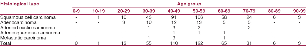 | Table 2: Histological classification and age distribution of cervical malignancies
Click here to view |
| Conclusion | | |
This study indicates that little has changed in the relative frequency of squamous cell carcinoma of the cervix in Benin City. However, there appears to be a slightly increased frequency in the diagnosis of adenocarcinoma of the cervix which may be attributed to the cervical cancer screening established about the same period and possibly increased awareness of glandular malignancies of the cervix. Since the frequency of cervical cancer is high, well-articulated regional and national policies should be put in place to increase detection of preinvasive lesions which in turn will decrease the frequency of cervical cancer to what obtains in developed countries.
| References | | |
| 1. | Global action against cancer. World Health Organization and International Union against cancer. World Health Organization; 2003.  |
| 2. | Parkin DM, Bray F, Ferlay J, Pisani P. Global cancer statistics, 2002. CA Cancer J Clin 2005;55:74-108. |
| 3. | Nofal M. Controlling cervical cancer in the developing world. IAEA Bull 1986:11-4. |
| 4. | Okobia MN, Aligbe JU. Pattern of malignant diseases at the University of Benin Teaching Hospital. Trop Doct 2005;35:91-2. |
| 5. | Quinn M, Babb P, Jones J, Allen E. Effect of screening on incidence of and mortality from cancer of cervix in England: Evaluation based on routinely collected statistics. BMJ 1999;318:904-8. |
| 6. | Aklimunnessa K, Mori M, Khan MM, Sakauchi F, Kubo T, Fujino Y, et al. Effectiveness of cervical cancer screening over cervical cancer mortality among Japanese women. Jpn J Clin Oncol 2006;36:511-8. |
| 7. | |
| 8. | World Health Organization. Tumours of the breast and female genital organs. Geneva: IARC Press; 2003. |
| 9. | Ijaiya MA, Aboyeji AP, Olatinwo AW, Buhari MO. Clinico-pathological presentation of primary cervical cancer seen in Ilorin, Nigeria. Niger J Surg Res 2002;4:89-93. |
| 10. | Ijaiya MA, Aboyeji PA, Buhari MO. Cancer of the cervix in Ilorin, Nigeria. West Afr J Med 2004;23:319-22. |
| 11. | Nwosu SO, Anya SE. Malignancies of the female genital tract at the University of Port Harcourt Teaching Hospital: A ten year review - 1990-1999. Niger Postgrad Med J 2004;11:107-9. |
| 12. | Olu-Eddo AN, Ekanem VJ, Umannah I, Onakevhor J. A 20 year histopathological study of cancer of the cervix in Nigerians. Nig Q J Hosp Med 2011;21:149-53. |
| 13. | Ogunbiyi JO. Epidemiology of Cancer in Ibadan: Tumours in adults. Arch Ib Cancer Med 1999;1:9-12. |
| 14. | Oguntayo O, Zayyan M, Kolawole A, Adewuyi S, Ismail H, Koledade K. Cancer of the cervix in Zaria, Northern Nigeria. Ecancermedicalscience 2011;5:219. |
| 15. | Bassey EA, Ekpo MD, Abasiatai A. Female genital tract malignancies in Uyo, South-South Nigeria. Niger Postgrad Med J 2007;14:134-6. |
| 16. | Nkyekyer K. Pattern of gynaecological cancers in Ghana. East Afr Med J 2000;77:534-8. |
| 17. | Jeebun N, Agnihotri S, Manraj SS, Purwar B. Study of cervical cancers in mauritius over a twelve years period (1989-2000) and role of cervical screening. Vol. 3. Internet J Oncol 2005. |
| 18. | Ghouth AS, Bafageer SS. The pattern and distribution of malignancies reported in Hadramout, Yemen-2006. J Pak Med Assoc 2009;59:774-8. |
| 19. | Sen U, Sankaranarayanan R, Mandal S, Ramanakumar AV, Parkin DM, Siddiqi M. Cancer patterns in eastern India: The first report of the Kolkata cancer registry. Int J Cancer 2002;100:86-91. |
| 20. | Ahmad Z, Azad NS, Yaqoob N, Husain A, Ahsan A, Khan AN, et al. Frequency of primary solid malignant neoplasms in both sexes, as seen in our practice. J Ayub Med Coll Abbottabad 2007;19:53-5. |
| 21. | CancerStats, Ovarian Cancer. UK: Cancer Research, UK; 2011. p. 1-3. |
| 22. | O′Leary M, Sheaffer J, Finklestein J, Olshan A, Brown J. Female Genital Tract Cancer. SEER AYA Monograph. Ch. 14. National Cancer Institute. p. 164-72. |
| 23. | Jamal S, Mamoon N, Mushtaq S, Luqman M, Moghal S. The pattern of gynecological malignancies in 968 cases from Pakistan. Ann Saudi Med 2006;26:382-4. [ PUBMED]  |
| 24. | Ikechebelu JI, Onyiaorah IV, Ugboaja JO, Anyiam DC, Eleje GU. Clinicopathological analysis of cervical cancer seen in a tertiary health facility in Nnewi, South-East Nigeria. J Obstet Gynaecol 2010;30:299-301. |
[Table 1], [Table 2]
|


 Search Pubmed for