|
 
 |
| ORIGINAL ARTICLE |
|
| Year : 2013 | Volume
: 1
| Issue : 2 | Page : 66-69 |
|
Prevalence of dental anomalies in pretreatment orthodontic patients in Western Maharashtra, India: An epidemiological study
Aniket H Vibhute1, Nupura A Vibhute2, Rajendra Daule3
1 Department of Orthodontia, Bharati Vidyapeeth Dental College and Hospital, Pune, Maharashtra, India
2 Department of Oral Pathology and Microbiology, School of Dental Sciences, Krishna Institute of Medical Sciences, Deemed University, Karad, Maharashtra, India
3 Department of Conservative Dentistry, Bharati Vidyapeeth Dental College and Hospital, Pune, Maharashtra, India
| Date of Web Publication | 7-Aug-2013 |
Correspondence Address:
Nupura A Vibhute
C/O Dr. H.G. Vibhute, Opposite Hotel Greenfield, Uttekaranagar, Sadar Bazar, Satara, Maharashtra - 415 001
India
 Source of Support: Prevalence of dental Anomalies in Western Maharashtra, India, Conflict of Interest: None  | Check |
DOI: 10.4103/2321-3825.116286

Aim: The aim of this study was the evaluation of the prevalence and distribution of dental anomalies in the pre-treatment records of orthodontic patients at a rural dental OPD in Western Maharashtra, India. Materials and Methods: Retrospective examination was done of 220 panoramic radiographs, study models, and pre-orthodontic records. Dental anomalies were recorded using panoramic radiographs and study models and reported as descriptive statistics. Statistical Analysis Used: Descriptive analyses-using the Statistical Package for Social Science (SPSS 9.0)-were used. Results: Patients were between 8 and 14 years of age (mean 11.6 years). The patient types included: Class I: 123 (55.9%), Class II: 94 (42.7%), Class III: 17 (7.7%), and Superclass I: 6 (2.7%). Crowding and spacing were found in 162 (73.6%) and 40 (18.1%) of patients, respectively. It was found that 27.7% of patients had at least one dental anomaly: hypodontia being the most common 23 (10.4%), followed by microdontia 17 (7.7%), hyperdontia 14 (6.3%), transposition 9 (4.1%), root dilaceration 9 (4.1%), macrodontia 7 (3.2%), Talon's cusp 3 (1.4%), and fusion 2 (0.9%). Conclusions: The present study investigating the prevalence of various dental anomalies in orthodontic patients found that 27.7% of the patients showed at least one dental anomaly. No significant association between the occurrences of dental anomalies was found in the study. Prevalence and distribution of some dental anomalies in rural Indian orthodontic patients differed from other studies. Careful prior detection of dental anomalies would simplify orthodontic treatment plan and reduce complications. Keywords: Dental anomalies, malocclusion, orthodontics
How to cite this article:
Vibhute AH, Vibhute NA, Daule R. Prevalence of dental anomalies in pretreatment orthodontic patients in Western Maharashtra, India: An epidemiological study. J Orthod Res 2013;1:66-9 |
How to cite this URL:
Vibhute AH, Vibhute NA, Daule R. Prevalence of dental anomalies in pretreatment orthodontic patients in Western Maharashtra, India: An epidemiological study. J Orthod Res [serial online] 2013 [cited 2017 Apr 7];1:66-9. Available from: http://www.jorthodr.org/text.asp?2013/1/2/66/116286 |
| Introduction | |  |
Developmental anomalies of the dentition are found in a number of malocclusion patients. Anomalies in tooth number, shape, and position may lead to disturbances in maxillary and mandibular arch length and occlusion. These play a role in orthodontic treatment planning. Recently, studies have been conducted to find the prevalence of these anomalies in orthodontic patients.
In 1959, Lind [1] examined 1717 Swedish orthodontic patients and found that 3.6% had supernumerary teeth. Rose [2] did a survey of the incidence of congenitally missing teeth in 6000 orthodontic patients aged 7 to 14 years and found 4.3% had at least one congenitally missing tooth. A pilot twin study by Kotsomitis et al. [3] on 202 orthodontic patients (101 pairs) reported a prevalence of 29.7% for ectopic eruption and 8.4% for agenesis. Ben-Bassat and Brin [4] found that multiple congenitally missing teeth affected the skeletal pattern. Endo et al. [5] reported the association of hypodontia and craniofacial morphology in Japanese orthodontic patients.
Dental anomalies are encountered commonly in orthodontic patients. This study was aimed at evaluating their prevalence and distribution in a rural setting in western Maharashtra, India, to assay their role in orthodontic treatment planning to help in reducing complications in future.
| Materials and Methods | | |
Pre-orthodontic study models and panoramic radiographs of 220 subjects (98 males and 122 females) orthodontic patients from a private rural orthodontic OPD in a rural setting in western Maharashtra, India were retrospectively evaluated. Detailed medical, dental, and family histories were obtained for all subjects. The selection criteria are followed:
- No significant medical history
- No history of extraction or previous orthodontic treatment
- No craniofacial anomalies and syndromes
- Availability of good quality of study models and OPG
Method of Analysis of Dental Abnormalities
Panoramic radiographs and study models were examined for dental anomalies
The following dental anomalies were assessed:
- Number abnormalities (hypodontia and hyperdontia);
- Size abnormalities (macrodontia and microdontia);
- Shape abnormalities (fusion, gemination, Talon's cusp);
- Location abnormalities (transposition of tooth);
- Root abnormalities: dilaceration.
Data collected were pooled and analyzed for frequency and sex distribution.
| Results | | |
In this study retrospective examination was done of 220 panoramic radiographs, study models, and pre-orthodontic records.
In the present study, the sample group comprised 98 males (44.5%) and 122 females (55.5%).
Age ranged between 8 and 14 years of age (mean 11.6 years).
The patients were grouped in to: Class I: 123 (55.9%), Class II: 94 (42.7%), Class III: 17 (7.7%), and Superclass I: 6 (2.7%). Crowding and spacing problems were found in 162 (73.6%) and 40 (18.1%) of patients, respectively. The prevalence and distribution of dental anomalies: Of 220 patients, 159 (72.3%) showed no dental anomaly, whereas the rest 61 (27.7%) of patients had at least one dental anomaly.
Macrodontia and Microdontia
The percentage of macrodontia and microdontia in our study was 3.1 and 7.7%, respectively. The most common microdontia was of the maxillary lateral incisor.
Fusion, Gemination, Talon's cusp
In our study, fusion occurred in only 0.9% and no gemination was found. In our study, Talons's cusp was found in 1.4% cases.
Transposition of tooth
Our study found that the most frequently transposed teeth were the maxillary canine-lateral incisors. The prevalence of patients with ectopic eruption of teeth in this study was 4.1%.
The maxillary canine was the most common ectopic tooth.
Root dilaceration
Root dilaceration requires radiographic examination. It was observed in 4.1% of the patients in this study.
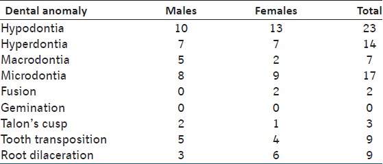
Frequencies of dental anomalies and sex distribution are shown in [Table 1].
Hypodontia
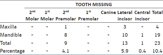
In our study, the most commonly missing tooth, excluding the third molar, was the mandibular lateral incisor 10(4.5%), followed by the mandibular second premolar 8 (3.6%) and the maxillary lateral incisor 3 (1.4%). No significant sex difference was found. Prevalence and incidence of hypodontia in our study are shown in [Table 2].
Hyperdontia
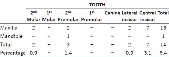
Our study found mesiodens in 3.1% of orthodontic patients. The other commonly occurring supernumerary teeth in our study were maxillary second premolar 1.4% and both maxillary second molar and mandibular lateral incisor appearing in 0.9%. [Table 3] shows the prevalence and incidence of hyperdontia in our study.
| Discussion | | |
Although there have been several studies reporting the prevalence of various dental anomalies, no reported study has been conducted on Indian rural orthodontic patients. In the present study, the prevalence of permanent tooth anomalies in patients who underwent orthodontic treatment was analyzed providing an estimation of the prevalence of dental anomalies in Indian rural orthodontic patients as a whole.
Hypodontia
Hypodontia means missing teeth. Multiple missing teeth not only cause malocclusion but also make orthodontic treatment difficult due to poor occlusal support and stability. Some missing teeth have been reported in association with at least one other dental anomaly, [6],[7] and may complicate orthodontic problems. The most common missing tooth in orthodontic patients varies among the studied groups. Endo et al. [5] reported that the most commonly affected tooth was in the mandibular second premolar. The maxillary lateral incisor was the most frequent in many studies. [8],[9],[10] Findings in our study showed similar results, as the most commonly missing tooth, excluding the third molar, was the mandibular lateral incisor 10 (4.5%), followed by the mandibular second premolar 8 (3.6%), and the maxillary lateral incisor 3 (1.4%). No significant sex difference was found.
Hyperdontia
Hyperdontia is increased number of teeth. In case of hyperdontia, the orthodontist plays a key role in the diagnosis and therapy through a comprehensive examination. The prevalence of supernumerary teeth is usually lower than that of tooth agenesis. [11] The prevalence of supernumerary teeth in orthodontic patients ranges between 0.3 and 1.37%. The most common site of supernumerary teeth is in the maxillary anterior region. [12]
Our study found mesiodens in 3.1% of orthodontic patients. Mesiodens may cause delay or ectopic eruption of the permanent incisor or further alter the occlusion and appearance. Early diagnosis is therefore needed for appropriate treatment; thereby reducing the invasiveness of surgery, orthodontic treatment, and possible complications. [13] Most of the studies reported mesiodens in terms of supernumerary teeth.
Macrodontia and Microdontia
Macrodontia, i.e. large teeth contribute to crowding and microdontia, i.e. small teeth cause spacing. Macrodontia is very much less common than microdontia. Compared with other studies [8],[9] the percentage of macrodontia and microdontia in our study was 3.1 and 7.7%, respectively. The most common microdontia was of the maxillary lateral incisor. [8],[9] The treatment of microdontia may require prosthetic treatment along with orthodontic treatment.
Fusion, Gemination, and Talon's cusp
These are though the rare anomalies that require management for esthetic reasons, caries control, and occlusal accomodation. Fusion and gemination in the general population are reportedly very low (0.19 and 0.22%, respectively) and extremely limited in orthodontic reports. No gemination was found in our study. Altug-Atac and Erdem [9] reported the frequency of fusion and gemination was 0.23 and 0.07%, respectively. The findings in our study were comparable with these studies with fusion and study Talons's cusp occurring in only 0.9 and 1.4% cases, respectively.
Transposition of tooth
Teeth transposition is an eruption anomaly that involves the permanent dentition (incidence 0.3-0.4%). [14],[15] Transposition are more frequently seen in the maxilla, [16] as in our study, and affecting (in descending order) the canines and first premolars, the canines and lateral incisors and the lateral and central incisors. [16],[17] Our study found that the most frequently transposed teeth were the maxillary canine-lateral incisors. Transposition may occur with other anomalies, such as aplasia, peg-shaped lateral incisor, and deciduous teeth retention. [14] Diagnosis could be made at the radiological level.
The prevalence of patients with ectopic eruption of teeth in this study was 4.1%. This is little less than the incidence of 7.2% reported by Bergstrom [18] who examined panoramic radiographs of 2589 school children between the age of 8 and 9 years. The maxillary canine was the most common ectopic tooth, confirming the results of previous investigators. [8],[19]
Root dilaceration
Root dilaceration requires radiographic examination. It was observed in 4.1% of the patients in this study. Panoramic radiography alone is not the method of choice for diagnosis of root dilaceration which can occur in a labial or lingual direction and may not be detected by panoramic radiography. Additional radiographs from different angles would be more useful for the diagnosis of this type of anomaly.
| Conclusion | | |
The present study investigated the prevalence of various dental anomalies in orthodontic patients. It was found that 27.7% of the patients showed at least one dental anomaly. Hypodontia was the most prevalent dental anomaly. Prevalence of dental anomalies was higher for second premolars and lateral incisors. No significant association between the occurrence of dental anomalies and sex distribution was found. Prevalence and distribution of some dental anomalies in rural Indian orthodontic patients differed from other studies. Orthodontists should concern about the difference in dental anomalies in various group of patients. Careful diagnosis would simplify treatment plan and reduce complications.
| References | | |
| 1. | Lind V. Medfodda antalsvariationer i permanenta dentitionen. Odont Rev 1959;10:176-89. 
|
| 2. | Rose JS. A survey of congenitally missing teeth, excluding third molars, in 6000 orthodontic patients. Dent Pract (Bristol) 1996;17:107-14.
|
| 3. | Kotsomitis N, Dunne MP, Freer TJ. A genetic aetiology for some common dental anomalies: A pilot twin study. Aust Orthod J 1996;14:172-8.
[PUBMED] |
| 4. | Ben-Bassat Y, Brin I. Skeletodental patterns in patients with multiple congenitally missing teeth. Am J Orthod Dentofacial Orthop 2003;124:521-5.
[PUBMED] |
| 5. | Endo T, Ozoe R, Kubota M, Akiyama M, Shimooka S. A survey of hypodontia in Japanese orthodontic patients. Am J Orthod Dentofacial Orthop 2006;129:29-35
|
| 6. | Gomes R, da Fonseca J, Paula L, Faber J, Acevedo A. Prevalence of hypodontia in orthodontic patients in Brasilia, Brazil. Eur J Orthod 2010;32:302-6.
|
| 7. | Garib D, Peck S, Gomes S. Increased occurrence of dental anomalies associated with second-premolar agenesis. Angle Orthod 2009;79:436-41.
|
| 8. | Uslu O, Akcam O, Evirgen S, Cebeci L. Prevalence of dental anomalies in various malocclusions. Am J Orthod Dentofacial Orthop 2009;135:328-5.
|
| 9. | Altug-Atac AT, Erdem D. Prevalence and distribution of dental anomalies in orthodontic patients. Am J Orthod Dentofacial Orthop 2007;131:510-4.
[PUBMED] |
| 10. | Silva Meza R. Radiographic assessment of congenitally missing teeth in orthodontic patients. Int J Paediatr Dent 2003;13:112-6.
[PUBMED] |
| 11. | Legovic M, Ceranic I, Cehich A. Anomalies in the number of permanent teeth in orthodontic patients in 2 localities in Croatia. Schweizer Monatsschrift fur Zahnmedizin 1990;100:286-90.
|
| 12. | Gabris K, Fabian G, Kaan M, Rozsa N, Tarjan I. Prevalence of hypodontia and hyperdontia in paedodontic and orthodontic patients in Budapest. Community Dent Health 2006;23:80-2.
|
| 13. | Russell K, Folwarczna M. Mesiodens-diagnosis and management. J Can Dent Assoc 2003;69:362-6.
|
| 14. | Budai M, Ficzere I, Gabris K, Tarjan I. Frequency of transposition and its treatment at the Department of Pedodontics and Orthodontics of Semmelweis University in the last five years. Fogorvo Sz 2003;96:21-4.
|
| 15. | Yilmaz HH, Turkkahraman H, Sayin MO. Prevalence of tooth transpositions and associated dental anomalies in a Turkish population. Dentomaxillofac Radiol 2005;34:32-5.
|
| 16. | Ely N, Sherriff M, Cobourne M. Dental transposition as a disorder of genetic origin. Eur J Orthod 2006;28:145-1.
|
| 17. | Shaipura Y, Kuftinec M. Maxillary tooth transpositions: Characteristic features and accompanying dental anomalies. Am J Orthod Dentofacial Orthop 2001;119:127-34.
|
| 18. | Bergstr¨om K. An orthopantomographic study of hypodontia, supernumeraries and other anomalies in school children between the ages of 8-9 years. Swed Dent J 1977;1:145-57.
|
| 19. | Becker A, Smith P, Behar R. The incidence of anomalous maxillary lateral incisors in relation to palatally-displaced cuspids. Angle Orthod 1981;51:24-9.
[PUBMED] |
[Table 1], [Table 2], [Table 3]
|


 Search Pubmed for