|
 
 |
| ORIGINAL ARTICLE |
|
| Year : 2014 | Volume
: 2
| Issue : 1 | Page : 32-37 |
|
Real-time cell analysis of cytotoxicity of orthodontic cements on gingival fibroblasts
Firat Ozturk1, Ebubekir Toy1, Erdem Hatunoglu1, Buket S Bozkurt2, Sema S Hakki3
1 Department of Orthodontics, Faculty of Dentistry, Inonu University, Malatya, Turkey
2 Research Center of Faculty of Dentistry, Selcuk University, Konya, Turkey
3 Department of Periodontology, Faculty of Dentistry, Selcuk University, Konya, Turkey
| Date of Web Publication | 29-Jan-2014 |
Correspondence Address:
Ebubekir Toy
Department of Orthodontics, Faculty of Dentistry, Inonu University, 44280 Malatya
Turkey
 Source of Support: None, Conflict of Interest: None  | Check |
DOI: 10.4103/2321-3825.125922

Introduction: To evaluate the cytotoxicity of four different orthodontic cement materials using the real-time xCELLigence system. Materials and Methods: Four orthodontic glass ionomer cements (GICs) were selected for this study, namely: GC Fuji (GC Cooperation), Ultra Band Lok (Reliance), Multi Cure (3M Unitek), and Meron (Voco). Ten test cylinders (measuring 5 Χ 2 mm) of each material were fabricated, making a total of 40 cylinders. The samples were incubated in Dulbecco modified Eagle medium (DMEM) culture medium for 72 hours. Human gingival fibroblasts (HGFs) were maintained with DMEM containing 10% fetal bovine serum. A real-time cell analyzer (RT-CES, xCELLigence) was used to evaluate cell survival. After seeding 200 μL of the cell suspensions into the wells (10,000 cells/well), gingival fibroblasts were treated with bioactive components released from cement materials and were monitored every 15 minutes for a period of 88 hours. For proliferation experiments, the statistical analyses used were one-way analysis of variance (ANOVA) and Tukey-Kramer multiple comparisons tests. Results: When the data were evaluated at 24 and 48 hrs, all tested materials showed statistically significant decreases in HGF cell index compared to the control group (P < 0.001). Conclusion: According to the results of this study, all tested cements were found to have cytotoxic effects to the HGFs. Keywords: Cytotoxicity, human gingival fibroblasts, orthodontic cements, xCELLigence system
How to cite this article:
Ozturk F, Toy E, Hatunoglu E, Bozkurt BS, Hakki SS. Real-time cell analysis of cytotoxicity of orthodontic cements on gingival fibroblasts. J Orthod Res 2014;2:32-7 |
How to cite this URL:
Ozturk F, Toy E, Hatunoglu E, Bozkurt BS, Hakki SS. Real-time cell analysis of cytotoxicity of orthodontic cements on gingival fibroblasts. J Orthod Res [serial online] 2014 [cited 2018 Mar 27];2:32-7. Available from: http://www.jorthodr.org/text.asp?2014/2/1/32/125922 |
| Introduction | |  |
Several luting systems are routinely used in dentistry for the cementation of prosthetic restorations and orthodontic appliances. Conventional glass ionomer cements (GICs) are the most widespread materials since their introduction by Wilson and Kent, due to their ability to chemically adhere to mineralized tissue and metal. [1]
According to variations in chemical composition and setting reaction, these products have been categorized as resin-modified GICs (RMGICs) or modified composites, and used for cementing orthodontic bands. [2],[3] RMGICs are dual setting: upon mixing, the acid-base reaction occurs and the light-initiated free-radical polymerization of resin also occurs. [4] Polyacid-modified composite resins (PAMCR) are the composite materials, consisting of partially silanized ion-leachable glass embedded in a light-activated polymeric matrix. [5]
Orthodontic materials have to contact or interact with body tissue and fluids over extended periods. Orthodontic materials, such as brackets, wires, composites, and cements, consist of some compounds known to have allergic, cytotoxic, mutagenic, and/or carcinogenic potential. [6] Evaluating the cytotoxicity and biocompatibility of orthodontic materials is as important as evaluating their physiological or mechanical properties.
Triethylene-glycoldimethacrylate (TEGDMA), urethane dimethacrylate (UDMA), 2-hydroxyethylmethacrylate (HEMA), bisphenol A-diglycidyl dimethacrylate (Bis-GMA), and methyl methacrylate (MMA) are released from the orthodontic resin-based adhesives. The release of these ions and their diffusion through oral tissue has cytotoxic effects. [2],[7],[8],[9] Furthermore, consistent exposure to dental monomers could cause allergic dermatitis, drowsiness, headache, and anorexia. [10] Evaluation of the cytotoxicity of compounds that had been used in dental resin materials showed a relationship between their monomeric structure and the degree of cytotoxicity, since TEGDMA, and mainly Bis-GMA and UDMA, are highly cytotoxic. [11]
Although the development and improvements related to the GICs materials are very satisfying and promising, the cytotoxicity of these materials is still a controversial question for orthodontists. In addition, there is insufficient evidence from published studies related to the cytotoxicity of the commercially available orthodontic GICs at present. Orthodontists are using a large variety of bonding and banding adhesives that, insofar as it is possible, must be harmless. Newer orthodontic adhesive materials present new challenges because of their potential for interaction.
Different testing methods have been used to evaluate the cytotoxic effects of adhesives, including inhibition of cell growth, effects on membrane or cytoplasmic markers cytolysis, mitochondrial dehydrogenase of active cells (MTT), [12] and changes in metabolic activity. [13] The xCELLigence system allows observing the indices of cultured cells using electrical impedance. The continuous monitoring of the cultured cells by using specially designed microtiter plates and its interdigitated gold microelectrodes of this system enables it to indicate cell viability, cell death, and reduced proliferation. [14]
Few comprehensive data are available in orthodontic literature regarding the cytotoxicity of different types of the orthodontic GICs. [8] The xCELLigence system is a new technique used to test the cytotoxicity of dental materials. [15],[16],[17] Therefore, the aim of the present study was to evaluate the cytotoxic effects of four different type orthodontic GICs using the xCELLigence system.
| Materials and Methods | | |
The orthodontic GICs selected were GC Fuji (GC Cooperation), Ultra Band Lok (Reliance), Multi Cure (3M Unitek), and Meron (Voco). [Table 1] includes information about the contents and manufacturers of the tested cements.
As the test procedures for this investigation, we followed the recommendations of the ISO-standard 10993-5. The specimens were fabricated according to the manufacturers' recommendations in standard Teflon discs. All specimens were prepared 5 mm in diameter and 2 mm in thickness and handled under aseptic conditions to prevent the contamination of the cell culture tests. To minimize the oxygen inhibition and maximize the surface smoothness we prepared the test samples using Mylar and glass slabs. Afterwards, the samples were disinfected under UV light. Forty cylinders, measuring 5 × 2 mm, were fabricated consisting of ten samples of tested cements for cytotoxicity testing. The samples were immersed in 7 mL of culture medium for 24 hrs at 37°C to extract residual monomer or cytotoxic substances. The culture medium containing material extracts was sterile, filtered for use on the cell cultures as applied by Öztürk et al. [5]
Cell Culture
We isolated human gingival fibroblasts (HGF) from healthy samples of gingiva removed for crown lengthening purposes. All patients gave informed consent before providing the samples. This protocol was approved by Ethics Committee of the Selcuk University, Faculty of Dentistry. The procedure was that the gingival tissues were cut into small pieces, rinsed with biopsy media, placed in tissue culture dishes, and incubated in biopsy medium in a humidified atmosphere of 95% air and 5% CO 2 at 37°C overnight. The following day, the biopsy medium was replaced with a culture medium, (Dulbecco modified Eagle medium [DMEM] 10% fetal bovine serum, 100 units/mL penicillin, 100 μg/mL streptomycin). After the dish was completely covered by the cells, they were passaged with 0.25% trypsin and 0.1% ethylene diaminotetraacetic acid (EDTA). HGFs were used between the fourth and sixth passage for all experiments as applied by Öztürk et al., [15] Malkoc et al., [16] and Hakki and Bozkurt. [17]
Preparation of Materials
Ten cylinders of each material were left to set for 2 days at 37°C. Materials were incubated in DMEM culture medium (the surface area-to-volume ratio of the specimen to the cell-culture medium was 3 cm 2 /mL) for 72 hrs according to ISO 10993-5 standards. Gingival fibroblasts were maintained with DMEM containing 10% fetal bovine serum.
The xCELLigence system (Roche Applied Science, Mannheim, Germany, and ACEA Biosciences, San Deigo, Calif) is a relatively new technique to evaluate cytotoxicity of materials. According to the instrument operator's manual, this system consists of four main components: the impedance-based real-time cell analyzer (RTCA), the RTCA single plate station, the RTCA computer with integrated software, and disposable E-plate 96. The RTCA single plate station fits inside a standard tissue-culture incubator. To monitor and detect the physiologic changes of the cells on the electrodes, the system measured the electronic impedance of the sensor electrodes. Voltage of 20 mV was applied to the electrodes during the RTCA measurement. The impedance measured between electrodes in each well depends on electrode geometry, ion concentration in the well, and whether the cells are attached to the electrodes. If there are no cells, electrode impedance is mainly determined by the ion environment both at the electrode-solution interface and in the bulk solution. In the presence of cells, cells attached to the electrode sensor surfaces act as insulators and thereby alter the local ion environment at the electrode-solution interface, leading to increased impedance. Thus, electrode impedance has larger value when more cells are growing on the electrodes. The system loaded the data obtained from cell index units to Excel software (Microsoft, Seattle, Wash) for any type of analysis. [18],[19]
To evaluate cell survival, the xCELLigence system was used, according to the instructions of the supplier. After seeding 200 μL of the cell suspensions into the wells (10,000 cells/well) of the E-plate 96, gingival fibroblasts were treated with bioactive components released from cement materials and were monitored every 15 min for a period of 88 hours. [15],[16]
Statistical Analysis
All calculations were obtained using the RTCA-integrated software of the xCELLigence system. RTCA software performs a curve-fitting of a selected "sigmoidal dose-response equation" to the experimental data points. Data are represented as mean [mmol/L] SD (n = 5).
One-way analysis of variance (ANOVA) and Tukey-Krammer multiple comparison tests were used for proliferation experiments and gene expressions. The data are represented as mean and standard deviation. The level of significance was set as P < 0.05.
| Results | | |
For optimal concentration to cell proliferation and viability measurements we indicated that, 10,000 cells/well were added in the E-Plate 96.
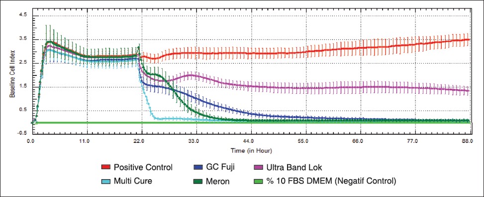
According to the ANOVA results, when the data were evaluated at 24 and 48 hrs, all tested materials showed statistically significant decreases in the HGF cell index compared to the control group (P < 0.001) [Figure 1].
According to our study's results, HGF cell proliferation decreased with time relatively. Although all four cements were found to decrease the cell viability significantly at 24 hrs, we observed differences among tested materials. Resin-modified glass ionomer cements (Multi cure, GC Fuji) showed lower value than the conventional GIC and PAMCR cements.
| Discussion | | |
The materials used in the oral cavity must be non-toxic, biocompatible, and have adequate mechanical properties. [20] Cytotoxicity of dental resins and their elutions have been shown in several studies. [21],[22] Thompson et al. [23] studied the amount of materials released from orthodontic adhesives and found that up to 14% of the total material could leach after 48 hours. Elution of residual unpolymerized monomers is a major cause of the cytotoxic effects. Therefore, the biocompatibility of these materials must be studied. Biocompatibility means that the tissues of the patient who comes into contact with the materials do not suffer from any toxic, irritating, inflammatory, allergic, mutagenic, or carcinogenetic action. [24] In the field of dentistry, orthodontics is the branch in which the problem of biocompatibility is most felt, since the patients are young and therefore more susceptible to developing inflammatory reactions. [25]
In comparison to animal experiments, testing of dental materials by using cell culture can be carried out easily, remade again or anew, controlled precisely, and is cost effective. [8] It has been reported that cytotoxicity experiments are appropriate initial tests, recommended to evaluate cytopathic effects caused by materials or their extracts, on the culture of cells. [26] Therefore, the aim of this study was to investigate the cytotoxic effects of different types of orthodontic cement using the xCELLigence system.
Cytotoxicity of dental products has been evaluated with inhibition of cell growth, effects on membrane or cytoplasmic markers cytolysis, mitochondrial dehydrogenase of active cells (MTT), and changes in metabolic activity. [13] In our study, we used the real-time xCELLingence system to investigate the cytotoxic effects of the adhesive compounds: UDMA, Bis-GMA, and HEMA on HGFs by continuous monitoring of the cell growth, proliferation, and viability.
The evaluation of cell proliferation, viability, and cytotoxicity, and even the physiological state of the cells can be obtained by real-time and continuous monitoring. The xCELLigence system also reduces expensive experimental subject usage in conventional cell analysis. During the experimental period information about cell growth, morphological changes, and cell death are detected with this system. Also, time-dependent physiological values, which can be more informative than the single-value endpoints of classical toxicity testing, can be precisely measured with this protocol. [15],[18] Real-time system has some advantages compared to conventional end-point cell-based assays, dynamic monitoring of cell response, especially cell adhesion, spreading, proliferation, and cell death for in vitro assays and also achieves both cell and assay conditions before and during the experiment. Cell reactions to a chemical exposure can be obtained in real time, which are not achieved by the MTT or other viability assays. [15],[27]
In humans, gingival fibroblasts are highly exposed to monomers or co-monomers after release from acrylic materials in the oral cavity. [28] Therefore, in this study, we used human gingival fibroblasts. Cement disks were prepared according to the ISO standards (10993-5) for the experiments; the cement was left to set in a dry atmosphere and to contact with air. However, the dental cements complete their setting in a wet environment clinically. Thinking that this method would be more analogous to the clinical conditions, we used different concentrations of MTA in DMEM containing 5% fetal bovine serum. Furthermore, the released bioactive components might interact with surrounding tissues differently. Our test method of cytotoxicity of cement components showed EC values for UDMA, Bis-GMA, or HEMA that are comparable to the end-point XTT-based viability assay data. [27]
For this experiment 10,000 cells/well were seeded in the E-Plate 96. Urcan et al. [18] investigated the cytotoxicity of the composites on HGFs and showed that the response seen in the 10,000-20,000 cells/well experiments reflects cell cycle effects, while the concentration of 40,000 cells/well was not suited for further experimentation, possibly because of a too-high cell density and the resulting contact inhibition.
According to our study's results, HGF cell proliferation decreased with time relatively. Although all four materials were found significantly cytotoxic at 24 hrs, we observed differences among the materials. Resin-modified glass ionomer cements (Multi cure, GC Fuji) were more cytotoxic than the conventional GIC and PAMCR cements. de Souza Costa et al. [7] examined the cytotoxic effects of GICs and RMGICs and stated that all experimental materials were cytotoxic to the odontoblast cells; the GICs were the least cytotoxic. Several in vitro studies assessed the cytotoxicity of conventional and RMGICs on cultured cells and showed that RMGICs had more intense cytotoxic effects than the conventional GICs. [2] RMGICs contain methacrylate monomers. For example, 3M Multi Cure cement contains HEMA; GC Fuji contains UDMA. The increased cytotoxicity of the RMGICs has been mainly attributed to the release of the monomers UDMA and HEMA, which are frequently added to their chemical composition because they act as both a consolvent and comonomer. [1] Methacrylate monomers such as HEMA and UDMA are incorporated in the lipid bilayers of cell membranes which are solubilized by the unreacted monomers. [29] This mechanism of action of uncured leached monomers on the cell membrane may be responsible for the high cytotoxicity of RMGICs observed in the present study.
When we evaluated the cytotoxicity of the cements at 48 hrs, all tested material showed cytotoxicity. PAMCR cement (Ultra Band Lok), which contains Bis-GMA, showed the least cytotoxic effects. Lee et al. [30] investigated the cytotoxicity of resin monomers using MTT. They stated that all experimental monomers exhibited a dose-dependent cytotoxic effect and ranked the cytotoxicity GMA>TEGDMA>HEMA. Studies have indicated that Bis-GMA, which is the main monomer eluted from dental composites, is the most potent toxic component among dimethacrylate derivatives. [31] This finding does not agree with the literature; this may be a result of the test method sensitivity. However, Urcan et al. [18] investigated the cytotoxicity effects of dental composites containing Bis-GMA, HEMA, TEGDMA, and UDMA on HGFs for a 24 hr exposure using the xCELLigence system, and they found that Bis-GMA had significantly higher cytotoxic effects compared to UDMA, TEGDMA, and HEMA. This result conflicted with the present study. We can speculate that these differences may depend on different ingredients' interactions with resin cements or composites.
The study detected potential toxic effects in orthodontic cements, which warrants further in vivo testing to reduce the potential cytotoxic effects. These cements have been generally used for band cementation, where the adhesives may come into intimate contact with the subgingival tissues in an orthodontic clinic. The clinician should use only as much material as necessary and should take care to remove excess cements.
In vitro cytotoxicity tests cannot be completely applied to clinical conditions. They clarify some of the biological effects of dental materials and their components by providing more details. [8] Cytotoxicity testing allows a comparison among available products and provides information to help choose a material with optimal conditions.
| Conclusion | | |
According to the results of the present study, we may conclude that all tested cements have cytotoxic effects to the HGFs. However, further studies using different test methods are needed.
| References | | |
| 1. | Bakopoulou A, Mourelatos D, Tsiftsoglou AS, Giassin NP, Mioglou E, Garefis P. Genotoxic and cytotoxic effects of different types of dental cement on normal cultured human lymphocytes. Mutat Res 2009;672:103-12. 
[PUBMED] |
| 2. | McCabe JF. Resin-modified glass-ionomers. Biomaterials 1998;19:521-7.
[PUBMED] |
| 3. | Uysal T, Ramoglu SI, Ertas H, Ulker M. Microleakage of orthodontic band cement at the cement-enamel and cement-band interfaces. Am J Orthod Dentofacial Orthop 2010;137:534-9.
|
| 4. | Rejman DJ, Eliades T, Bradley TG, Eliades G. Polymerization efficiency of glass-ionomer and resin adhesives under molar bands. Angle Orthod 2008;78:549-52.
[PUBMED] |
| 5. | Meyer JM, Cattani-Lorente MA, Dupuis V. Compomers: Between glass-ionomer cements and composites. Biomaterials 1998;19:529-39.
[PUBMED] |
| 6. | Beyersmann D, Hartwig A. Carcinogenic metal compounds: Recent insight into molecular and cellular mechanisms. Arch Toxicol 2008;82:493-512.
[PUBMED] |
| 7. | de Souza Costa CA, Hebling J, Garcia-Godoy F, Hanks CT. In vitro cytotoxicity of five glass-ionomer cements. Biomaterials 2003;24:3853-8.
[PUBMED] |
| 8. | Malkoc S, Corekci B, Botsali HE, Yalçin M, Sengun A. Cytotoxic effects of resin-modified orthodontic band adhesives. Are they safe? Angle Orthod 2010;80:890-5.
|
| 9. | Ulker HE, Sengun A. Cytotoxicity evaluation of self adhesive composite resin cements by dentin barrier test on 3D Pulp Cells. Eur J Dent 2009;3:120-6.
|
| 10. | Anderson RL, Stasior OG. Self-curing methyl methacrylate: Is it safe? Ophthalmic Surg 1976;7:28-30.
[PUBMED] |
| 11. | Altintas SH, Usumez A. Evaluation of monomer leaching from a dual cured resin cement. J Biomed Mater Res B Appl Biomater 2008;86:523-9.
[PUBMED] |
| 12. | Ulker M, Ulker HE, Zortuk M, Bulbul M, Tuncdemir AR, Bilgin MS. Effects of current provisional restoration materials on the viability of fibroblasts. Eur J Dent 2009;3:114-9.
|
| 13. | Hensten-Pettersen A. Comparison of the methods available for assessing cytotoxicity. Int Endod J 1988;21:89-99.
[PUBMED] |
| 14. | Ke N, Wang X, Xu X, Abassi YA. The xCELLigence system for real-time and label-free monitoring of cell viability. Methods Mol Biol 2011;740:33-43.
[PUBMED] |
| 15. | Öztürk F, Malkoc S, Ersöz M, Hakki SS, Bozkurt BS. Real-time cell analysis of the cytotoxicity of the components of orthodontic acrylic materials on gingival fibroblasts. Am J Orthod Dentofacial Orthop 2011;140:e243-9.
|
| 16. | Malkoç S, Öztürk F, Çörekçi B, Bozkurt BS, Hakki SS. Real-time cell analysis of the cytotoxicity of orthodontic mini-implants on human gingival fibroblasts and mouse osteoblasts. Am J Orthod Dentofacial Orthop 2012;141:419-26.
|
| 17. | Hakki SS, Bozkurt SB. Effects of different setting of diode laser on the mRNA expression of growth factors and type I collagen of human gingival fibroblasts. Lasers Med Sci 2011;27:325- 31.
[PUBMED] |
| 18. | Urcan E, Haertel U, Styllou M, Hickel R, Scherthan H, Reichl FX. Real-time xCELLigence impedance analysis of the cytotoxicity of dental composite components on human gingival fibroblasts. Dent Mater 2010;26:51-8.
[PUBMED] |
| 19. | Roche Diagnostics. Introduction of the RTCA SP instrument. RTCA SP instrument operator′s manual. Indianapolis, Indiana: ACEA Biosciences; 2008. p. 14-6.
|
| 20. | Favero L, Brollo P, Bressan E. Orthodontic anchorage with specific fixtures: Related study analysis. Am J Orthod Dentofacial Orthop 2002;122:84-94.
[PUBMED] |
| 21. | Ahrari F, Tavakkol Afshari J, Poosti M, Brook A. Cytotoxicity of orthodontic bonding adhesive resins on human oral fibroblasts. Eur J Orthod 2010;32:688-92.
[PUBMED] |
| 22. | Annunziata M, Aversa R, Apicella A, Annunziata A, Apicella D, Buonaiuto C, et al. In vitro biological response to a light-cured composite when used for cementation of composite inlays. Dent Mater 2006;22:1081-5.
[PUBMED] |
| 23. | Thompson LR, Miller EG, Bowles WH. Leaching of unpolymerized materials from orthodontic bonding resin. J Dent Res 1982;61:989-92.
[PUBMED] |
| 24. | Jacobsen N, Hensten-Pettersen A. Occupational health problems and adverse patient reactions in orthodontics. Eur J Orthod 1989;11:254-64.
[PUBMED] |
| 25. | Grimsdottir MR, Hensten-Pettersen A, Kullmann A. Proliferation of nickel-sensitive human lymphocytes by corrosion products of orthodontic appliances. Biomaterials 1994;15:1157-60.
|
| 26. | Cao T, Saw TY, Heng BC, Liu H, Yap AU, Ng ML. Comparison of different test models for the assessment of cytotoxicity of composite resins. J Appl Toxicol 2005;25:101-8.
[PUBMED] |
| 27. | Reichl FX, Esters M, Simon S, Seiss M, Kehe K, Kleinsasser N, et al. Cell death effects of resin-based dental material compounds and mercurials in human gingival fibroblasts. Arch Toxicol 2006;80:370-7.
[PUBMED] |
| 28. | Wan Q, Rumpf D, Schricker SR, Mariotti A, Culbertson BM. Influence of hyperbranched multi-methacrylates for dental neat resins on proliferation of human gingival fibroblasts. Biomacromolecules 2001;2:217-22.
[PUBMED] |
| 29. | Fujisawa S, Kadoma Y, Komoda Y. 1H and 13C NMR studies of the interaction of eugenol, phenol, and triethyleneglycol dimethacrylate with phospholipid liposomes as a model system for odontoblast membranes. J Dent Res 1988;67:1438-41.
[PUBMED] |
| 30. | Lee DH, Lim BS, Lee YK, Ahn SJ, Yang HC. Involvement of oxidative stress in mutagenicity and apoptosis caused by dental resin monomers in cell cultures. Dent Mater 2006;22:1086-92.
[PUBMED] |
| 31. | Issa Y, Watts DC, Brunton PA, Waters CM, Duxbury AJ. Resin composite monomers alter MTT and LDH activity of human gingival fibroblasts in vitro. Dent Mater 2004;20:12-20.
[PUBMED] |
[Figure 1]
[Table 1]
|


 Search Pubmed for