
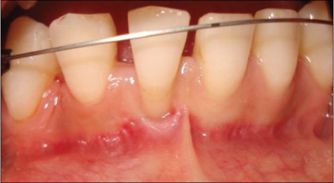
Figure 6: A Miller Class 2 gingival recession at tooth #41. Note the absence of mid-buccal keratinized tissue. Prior to anterior bracket placement after canine distalization
| Close | |
|
|
|
|
Figure 6: A Miller Class 2 gingival recession at tooth #41. Note the absence of mid-buccal keratinized tissue. Prior to anterior bracket placement after canine distalization
|
|