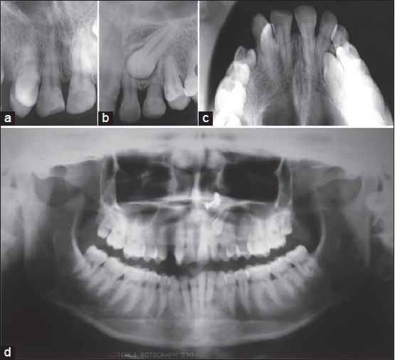
Figure 2: Pretreatment radiographs. (a) Intra oral periapical (IOPA) X-ray showed complete transposition of maxillary right canine and lateral incisor. (b) IOPA X-ray showed impacted maxillary left canine and retained deciduous canine. (c and d) Occluasal and orthopantomogram radiographs showed complete transposition of maxillary right canine and lateral incisor and impacted maxillary left canine