|


 |
| CASE REPORT |
|
| Year : 2012 | Volume
: 4
| Issue : 1 | Page : 43-45 |
|
|
A rare case of chondromyxoid fibroma mimicking spina ventosa
Kalyan Khan, Arghya Bandyopadhyay
Department of Pathology, North Bengal Medical College, Sushrutanagar, Darjeeling, India
| Date of Web Publication | 5-Sep-2012 |
Correspondence Address:
Kalyan Khan
Assistant Professor, Flat No. 11, 'BELA' Apartment, Netaji Subhas Road, Subhaspally, Siliguri, Darjeeling - 734 001
India
 Source of Support: None, Conflict of Interest: None
DOI: 10.4103/2006-8808.100354

 Abstract Abstract | | |
Chondromyxoid fibroma (CMF) is perhaps the rarest of all bone tumors. Classically it occurs in the metaphyseal region of the long bones surrounding the knee. The small bones of the feet are also commonly involved. But CMF occurring in small bones of the hand, however, is very uncommon. Tuberculous dactylitis is referred to as spina ventosa. The bones of the hands are more frequently affected than bones of the feet. We present a rare case of CMF occurring in the middle phalanx of the left middle finger which was misdiagnosed as spina ventosa clinicoradiologically. It can be a common mistake especially in areas where prevalence of tuberculosis is still high. This case once again stresses the need for biopsy and to consider CMF as a rare differential diagnosis in all suspected cases of spina ventosa. Keywords: Chondromyxoid fibroma, spina ventosa, tubercular dactylitis
How to cite this article:
Khan K, Bandyopadhyay A. A rare case of chondromyxoid fibroma mimicking spina ventosa. J Surg Tech Case Report 2012;4:43-5 |
How to cite this URL:
Khan K, Bandyopadhyay A. A rare case of chondromyxoid fibroma mimicking spina ventosa. J Surg Tech Case Report [serial online] 2012 [cited 2016 Jun 10];4:43-5. Available from: http://www.jstcr.org/text.asp?2012/4/1/43/100354 |
| Introduction | |  |
Chondromyxoid fibroma (CMF) is a rare benign cartilaginous bone tumor and perhaps the rarest of all bone tumors. [1] Classically it occurs in the metaphyseal region of the long bones surrounding the knee, but also found with relative frequency in other long bones, the pelvis, ribs, and small foot bones. The small bones of the hand, however, are very rarely involved. [2] Since 1948 till 1986 only six cases out of 136 reported cases of chondromyxoid fibroma were found to involve the hand. [3]
The radiographic appearances of CMF are those of a single, lytic lesion with lobulated margins, septations, cortical expansion, and a sclerotic rim but no periosteal reactions. [4] These features are similar to those of tubercular dactylitis of short bones of the hand which is referred to as spina ventosa. [5],[6] The common differential diagnoses of spina ventosa include osteomyelitis, metabolic diseases like gout, sarcoidosis, and tumors. [7],[8]
We present a case of histopathologically proven CMF occurring in the unusual site of middle phalanx of the left middle finger which was clinicoradiologically misdiagnosed as spina ventosa.
| Case Report | | |
A 21-year-old male patient presented with a slightly painful, slow growing, and hard, fusiform swelling of the left middle finger for the last 3 months. There was no history of fever and other routine investigations revealed only mild normocytic normochromic anemia and slightly raised erythrocyte sedimentation rate. On radiography, at the base of the middle phalanx of the left middle finger a lytic, radiolucent, lobulated lesion was seen with cortical expansion, a sclerotic rim, and septations. No calcification or periosteal reaction was noted [Figure 1]. In view of the high prevalence of tuberculosis in the region an initial clinicoradiological diagnosis of tubercular dactylitis of short bones of the hand which is referred to as spina ventosa was made. However, tuberculin test, chest radiogram, sputum for acid fast bacilli, and serum adenosine deaminase revealed negative results. Attempts of aspiration of the lesion yielded inadequate material. Hence, curettage was performed. On gross pathological examination fragments of gray-white lobulated mass with heterogeneous cut-surface was seen.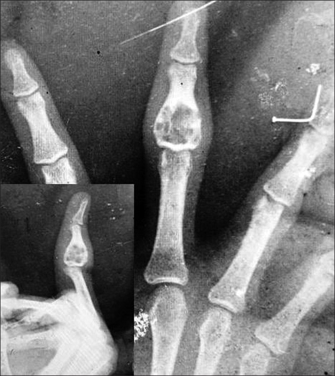 | Figure 1: X‑ray showing a lytic, radiolucent, lobulated lesion with
cortical expansion, a sclerotic rim, and septations at the base of the
middle phalanx of the left middle finger. No calcification or periosteal
reaction is noted
Click here to view |
Histopathology revealed a tumor having a lobular pattern of growth of spindle shaped and stellate cells, with abundant myxoid and chondroid intercellular material [Figure 2]. The lobules had a hypocellular center and a hypercellular periphery with scattered benign multinucleated giant cells and hemosiderin-laden macrophages [Figure 3]. One portion of the tumor showed pleomorphic large bizarre cells with irregular-shaped nuclei and a smudgy chromatin pattern indicative of pseudomalignant features [Figure 4]. The lamellar bony trabeculae showed no significant pathology [[Figure 2] inset]. A final diagnosis of chondromyxoid fibroma was made. | Figure 2: Tumor having a lobular pattern of growth of spindle shaped
and stellate cells, with abundant myxoid and chondroid intercellular
material; H and E ×100 (inset: Lamellar bony trabeculae showing no
significant pathology; ×100)
Click here to view |
 | Figure 3: The tumor lobules having a hypocellular center and a
hypercellular periphery with scattered benign multinucleated giant cells
(inset; H and E ×400) and hemosiderin‑laden macrophages; H and E ×100
Click here to view |
 | Figure 4: One portion of the tumor showing cells with pseudomalignant
features; H and E ×100 (inset; H and E ×400)
Click here to view |
Excisional curettage along with bone grafting was done and the patient is doing well 2 months after the treatment.
| Discussion | | |
Chondromyxoid fibroma is a rare benign tumor of chondral origin. The diagnosis is difficult, but as Jaffe emphasized, "its recognition is of some importance in that pathologically it may be mistaken for sarcoma and, as such, treated more radically than is necessary." [9] They may be encountered in any skeletal bones, mainly at the age of 20-30 years. The small bones of the hand, however, are rarely involved. [2],[10]
The radiographic appearances are those of a single, lytic lesion with lobulated margins, septations, cortical expansion, and a sclerotic rim. [4],[11] Magnetic resonance imaging (MRI) features are nonspecific for chondromyxoid fibroma. [1]
The possibility of chondromyxoid fibroma should always be considered when a focal bone lesion is evaluated that has geographic bone destruction, a sclerotic rim, lobulated margins, and septations. The diagnosis of chondromyxoid fibroma is most likely when the patient is in the second decade of life. [12]
On gross pathology, chondromyxoid fibroma is usually seen as a gray-white lobulated mass. The classic histological feature of a chondromyxoid fibroma is stellate or spindle-shaped cells arranged in lobules in a myxoid or chondroid background. [4],[11] The lobules are zonated. At the lobule periphery, the cells are longer, more numerous, and closer together. The cells are more stellate, thinner, less numerous, and farther apart in the center of the lobules. In a lobulated myxoid chondrosarcoma, which is a close differential diagnosis, the lesion shows liquefactive changes of the matrix, clear-cut permeation of surrounding bone, malignant radiographic features and most importantly hypercellularity throughout the lobules. In about half of the cases of CMF, giant cells are noted at the edge of the lobules. Bizarre pseudomalignant nuclei may be seen in nearly 20% of cases. [12],[13]
Tuberculous involvement of the metacarpals and phalanges is a rare presentation of extrapulmonary tuberculosis and extrapulmonary manifestations of tuberculosis have become increasingly important in the era of HIV/AIDS. [14]
The radiographic features of cystic expansion of the short tubular bones have led to the name of "spina ventosa" being given to tuberculous dactylitis of the short bones of the hand. The definite diagnosis of tuberculous dactylitis rests on bacteriological and histological studies. [7]
The present case once again emphasizes the need for histological and bacteriological confirmation of clinicoradiologically suspected cases of "spina ventosa" even in areas where tuberculosis is still highly prevalent. Simultaneously, CMF should be considered as a rare differential diagnosis in all suspected cases of spina ventosa especially if the patient is in the second decade of life.
| Acknowledgments | | |
The authors thank Dr. Goutam Bhattacharya, MS, Professor and Head, Department of Orthopaedics, North Bengal Medical College and Hospital. Dr. Sankar Kabiraj, MD, Professor and Head, Department of Radio diagnosis, North Bengal Medical College and Hospital.
| References | | |
| 1. | Yalniz E, Alicioglu B, Yalcin O, Yilmaz B. Non specific magnetic resonance features of chondromyxoid fibroma of the iliac bone. J BUON 2007;12:407-9. 
[PUBMED] |
| 2. | Slotcavage RL, Dickson BC, Ogilvie CM. Chondromyxoid fibroma involving the metacarpophalangeal joint. Orthopedics 2009;32:pii.
[PUBMED] |
| 3. | Anderson WJ, Bowers WH. Chondromyxoid fibroma of the proximal phalanx. A tumour that may be confused with chondrosarcoma. J Hand Surg Br 1986;11:144-6.
[PUBMED] |
| 4. | Rouas L, Malihy A, Cherradi N, Lamalmi N, Alhamany Z. Chondromyxoid fibroma of bone: A rare benign bone tumor in children. Rev Med Brux 2004;25:521-4.
[PUBMED] |
| 5. | Salimpour R, Salimpour P. Picture of the month. Tuberculous dactylitis. Arch Pediatr Adolesc Med 1997;151:851-2.
[PUBMED] |
| 6. | Zoga A, Lee VW. Paediatric case of the day. Tuberculosis dactylitis and primary pulmonary tuberculosis. AJR Am J Roentgenol 1999;173:815-7.
[PUBMED] |
| 7. | Jensen CM, Jensen CH, Paerregaard A. A diagnostic problem in tuberculous dactylitis. J Hand Surg Br 1991;16:202-3.
[PUBMED] |
| 8. | Sunderamoorthy D, Gupta V, Bleetman A. TB or not TB: An unusual sore finger. Emerg Med J 2001;18:490-1.
[PUBMED] |
| 9. | Jaffe HL, Lichtenstein L. Chondromyxoid fibroma of Bone: A distinctive benign tumor likely to be mistaken especially for chondrosarcoma. Arch Pathol 45:541-51.
|
| 10. | Semenova LA, Bulycheva IV. Chondromyxoid fibroma. Arkh Patol 2007;69:37-40.
[PUBMED] |
| 11. | Rouas L, Malihy A, Cherradi N, Lamalmi N, Alhamany Z. Chondromyxoid fibroma of bone: A rare benign bone tumor in children. Rev Med Brux 2004;25:521-4.
[PUBMED] |
| 12. | Wilson AJ, Kyriakos M, Ackerman LV. Chondromyxoid fibroma: Radiographic appearance in 38 cases and in a review of the literature. Radiology 1991;179:513-8.
[PUBMED] |
| 13. | Azorin D, Gil A, Sanchez-Aniceto G, Ballestin C, Martinez-Tello FJ. Chondromyxoid fibroma of the frontal sinus. Br J Oral Maxillofac Surg 2003;41:418-20.
|
| 14. | Agarwal S, Caplivski D, Bottone EJ. Disseminated tuberculosis presenting with finger swelling in a patient with tuberculous osteomyelitis: A case report. Ann Clin Microbiol Antimicrob 2005;3:4-18.
|
[Figure 1], [Figure 2], [Figure 3], [Figure 4]
|


