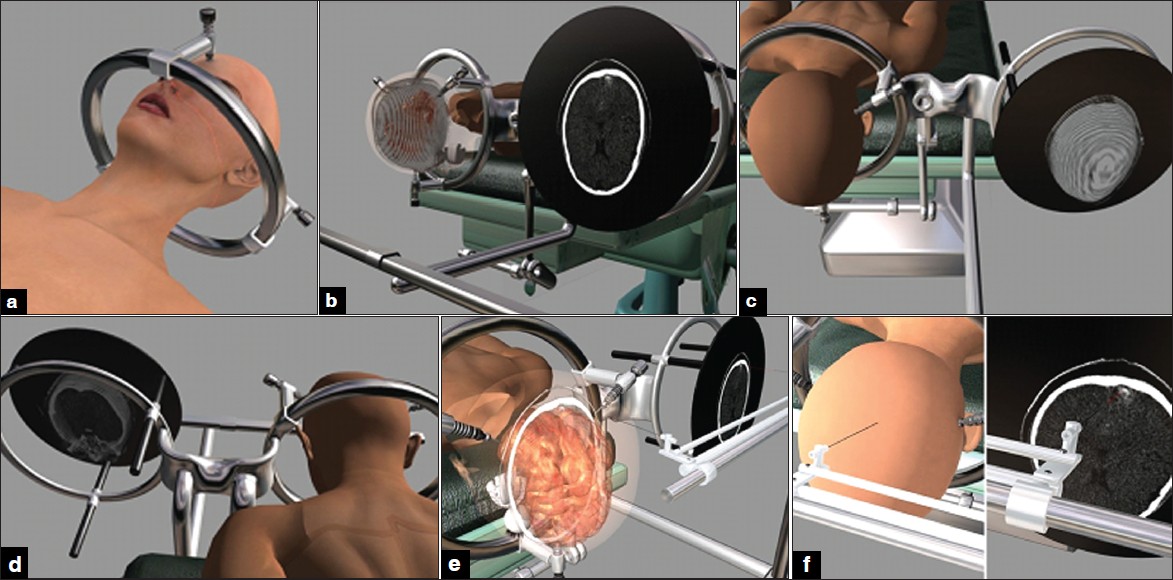
Figure 1: An occipital cavernous angioma at the tip of left occipital lobe. (a) The frame has been fi xed to the cranium. (b) The head of the patient have been fi xed to operating table at prone position and the images have been transferred to monitor. (c and d) The patient and images data have been adjusted. (e and f) The pantoghraph show that the data of the patient head and pathology are seen at the same location of their counterpart on LCD.